 Movie
Movie Controller


+ Open data

- Basic information
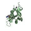
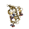
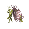
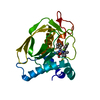
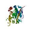
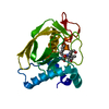
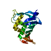
| Entry | Database: PDB / ID: 3rt2 | ||||||
|---|---|---|---|---|---|---|---|
| Title | Crystal structure of apo-PYL10 | ||||||
 Components Components | Abscisic acid receptor PYL10 | ||||||
 Keywords Keywords | HYDROLASE INHIBITOR / PYL10 / ABA-independent PP2C inhibitor / PP2Cs / abscisic acid / ABA receptor | ||||||
| Function / homology |  Function and homology information Function and homology informationabscisic acid binding / abscisic acid-activated signaling pathway / protein phosphatase inhibitor activity / signaling receptor activity / protein homodimerization activity / nucleus / plasma membrane / cytoplasm Similarity search - Function | ||||||
| Biological species |  | ||||||
| Method |  X-RAY DIFFRACTION / SYNCHROTRON / MOLECULAR REPLACEMENT / Resolution: 1.5 Å X-RAY DIFFRACTION / SYNCHROTRON / MOLECULAR REPLACEMENT / Resolution: 1.5 Å | ||||||
 Authors Authors | Hao, Q. / Yin, P. / Li, W. / Wang, L. / Yan, C. / Wang, J. / Yan, N. | ||||||
 Citation Citation | Journal: Mol.Cell / Year: 2011 Title: The Molecular Basis of ABA-Independent Inhibition of PP2Cs by a Subclass of PYL Proteins Authors: Hao, Q. / Yin, P. / Li, W. / Wang, L. / Yan, C. / Lin, Z. / Wu, J.Z. / Wang, J. / Yan, S.F. / Yan, N. | ||||||
| History |
|
- Structure visualization
| Structure viewer | Molecule: MolmilJmol/JSmol |
|---|
- Downloads & links
-Download
| PDBx/mmCIF format | 3rt2.cif.gz | 89.8 KB | Display | PDBx/mmCIF format |
|---|---|---|---|---|
| PDB format | pdb3rt2.ent.gz | 68.7 KB | Display | PDB format |
| PDBx/mmJSON format | 3rt2.json.gz | Tree view | PDBx/mmJSON format | |
| Others |  Other downloads Other downloads |
-Validation report
| Arichive directory | https://data.pdbj.org/pub/pdb/validation_reports/rt/3rt2ftp://data.pdbj.org/pub/pdb/validation_reports/rt/3rt2 | HTTPS FTP |
|---|
-Related structure data
| Related structure data | 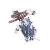 3rt0C  3kdhS C: citing same article ( S: Starting model for refinement |
|---|---|
| Similar structure data |


-Links
 PDBj
PDBj- Assembly
| Deposited unit | 
| |||||||||
|---|---|---|---|---|---|---|---|---|---|---|
| 1 |
| |||||||||
| 2 | 
| |||||||||
| Unit cell |
| |||||||||
| Components on special symmetry positions |
|
-Components
| #1: Protein | Mass: 20674.609 Da / Num. of mol.: 1 Source method: isolated from a genetically manipulated source Source: (gene. exp.)  |
|---|---|
| #2: Water | ChemComp-HOH /  Mass: 18.015 Da / Num. of mol.: 175 / Source method: isolated from a natural source / Formula: H2O Mass: 18.015 Da / Num. of mol.: 175 / Source method: isolated from a natural source / Formula: H2O |
| Has protein modification | Y |
-Experimental details
-Experiment
| Experiment | Method: X-RAY DIFFRACTION / Number of used crystals: 1 |
|---|
- Sample preparation
| Crystal | Density Matthews: 2.19 Å3/Da / Density % sol: 43.79 % |
|---|---|
| Crystal grow | Temperature: 291 K / Method: vapor diffusion, hanging drop / pH: 8 Details: 0.2M magnesium acetate, 20% PEG3350, pH 8.0, VAPOR DIFFUSION, HANGING DROP, temperature 291K |
-Data collection
| Diffraction | Mean temperature: 100 K |
|---|---|
| Diffraction source | Source: SYNCHROTRON / Site: SPring-8  / Beamline: BL41XU / Wavelength: 1 Å / Beamline: BL41XU / Wavelength: 1 Å |
| Detector | Type: RAYONIX MX225HE / Detector: CCD / Date: Jul 21, 2010 |
| Radiation | Monochromator: rotated-inclined double-crystal monochromator Protocol: SINGLE WAVELENGTH / Monochromatic (M) / Laue (L): M / Scattering type: x-ray |
| Radiation wavelength | Wavelength: 1 Å / Relative weight: 1 |
| Reflection | Resolution: 1.5→50 Å / Num. obs: 29479 / % possible obs: 99.7 % / Redundancy: 7.4 % / Biso Wilson estimate: 18.5 Å2 / Rmerge(I) obs: 0.04 |
| Reflection shell | Resolution: 1.5→1.55 Å / Redundancy: 7.3 % / Rmerge(I) obs: 0.378 / Mean I/σ(I) obs: 5.48 / Num. unique all: 2918 / % possible all: 100 |
- Processing
| Software |
| |||||||||||||||||||||||||||||||||||||||||||||||||||||||||||||||||||||||||||||
|---|---|---|---|---|---|---|---|---|---|---|---|---|---|---|---|---|---|---|---|---|---|---|---|---|---|---|---|---|---|---|---|---|---|---|---|---|---|---|---|---|---|---|---|---|---|---|---|---|---|---|---|---|---|---|---|---|---|---|---|---|---|---|---|---|---|---|---|---|---|---|---|---|---|---|---|---|---|---|
| Refinement | Method to determine structure: MOLECULAR REPLACEMENT Starting model: 3KDH Resolution: 1.5→28.598 Å / Occupancy max: 1 / Occupancy min: 0.35 / FOM work R set: 0.8697 / SU ML: 0.17 / σ(F): 0 / Phase error: 19.43 / Stereochemistry target values: ML
| |||||||||||||||||||||||||||||||||||||||||||||||||||||||||||||||||||||||||||||
| Solvent computation | Shrinkage radii: 0.83 Å / VDW probe radii: 1.1 Å / Solvent model: FLAT BULK SOLVENT MODEL / Bsol: 60.786 Å2 / ksol: 0.4 e/Å3 | |||||||||||||||||||||||||||||||||||||||||||||||||||||||||||||||||||||||||||||
| Displacement parameters | Biso max: 113.57 Å2 / Biso mean: 29.2402 Å2 / Biso min: 11.62 Å2
| |||||||||||||||||||||||||||||||||||||||||||||||||||||||||||||||||||||||||||||
| Refinement step | Cycle: LAST / Resolution: 1.5→28.598 Å
| |||||||||||||||||||||||||||||||||||||||||||||||||||||||||||||||||||||||||||||
| Refine LS restraints |
| |||||||||||||||||||||||||||||||||||||||||||||||||||||||||||||||||||||||||||||
| LS refinement shell | Refine-ID: X-RAY DIFFRACTION / Total num. of bins used: 10
|
