| Entry | Database: PDB / ID: 4p1n
|
|---|
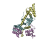
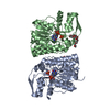
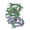
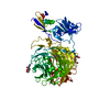
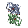
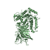
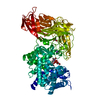
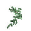
| Title | Crystal structure of Atg1-Atg13 complex |
|---|
 Components Components | |
|---|
 Keywords Keywords | PROTEIN TRANSPORT / Complex |
|---|
| Function / homology |  Function and homology information Function and homology information
histone H4S1 kinase activity / histone H2AS121 kinase activity / histone H3S57 kinase activity / histone H2BS14 kinase activity / histone H3S28 kinase activity / histone H2AS1 kinase activity / histone H3T6 kinase activity / histone H2BS36 kinase activity / ribosomal protein S6 kinase activity / histone H2AT120 kinase activity ...histone H4S1 kinase activity / histone H2AS121 kinase activity / histone H3S57 kinase activity / histone H2BS14 kinase activity / histone H3S28 kinase activity / histone H2AS1 kinase activity / histone H3T6 kinase activity / histone H2BS36 kinase activity / ribosomal protein S6 kinase activity / histone H2AT120 kinase activity / Rho-dependent protein serine/threonine kinase activity / AMP-activated protein kinase activity / eukaryotic translation initiation factor 2alpha kinase activity / Atg1/ULK1 kinase complex / 3-phosphoinositide-dependent protein kinase activity / histone H3T3 kinase activity / histone H3T45 kinase activity / DNA-dependent protein kinase activity / histone H2AXS139 kinase activity / phagophore assembly site membrane / protein localization to phagophore assembly site / autophagy of mitochondrion / piecemeal microautophagy of the nucleus / histone H3T11 kinase activity / histone H3S10 kinase activity / phagophore assembly site / reticulophagy / response to starvation / autophagosome assembly / mitophagy / autophagosome / regulation of autophagy / protein transport / non-specific serine/threonine protein kinase / protein serine kinase activity / ATP binding / cytosolSimilarity search - Function Helix Hairpins - #1900 / Autophagy-related protein 13, N-terminal / Autophagy-related protein 13 / Autophagy-related protein 13 / Serine/threonine-protein kinase Atg1-like, tMIT domain / : / Atg1-like, MIT domain 1 / ATG1-like, MIT domain 2 / Serine/threonine-protein kinase Atg1-like / HORMA domain superfamily ...Helix Hairpins - #1900 / Autophagy-related protein 13, N-terminal / Autophagy-related protein 13 / Autophagy-related protein 13 / Serine/threonine-protein kinase Atg1-like, tMIT domain / : / Atg1-like, MIT domain 1 / ATG1-like, MIT domain 2 / Serine/threonine-protein kinase Atg1-like / HORMA domain superfamily / Helix Hairpins / Helix non-globular / Special / Serine/threonine-protein kinase, active site / Serine/Threonine protein kinases active-site signature. / Protein kinase domain / Serine/Threonine protein kinases, catalytic domain / Protein kinase, ATP binding site / Protein kinases ATP-binding region signature. / Protein kinase domain profile. / Protein kinase domain / Protein kinase-like domain superfamilySimilarity search - Domain/homology |
|---|
| Biological species |  Kluyveromyces marxianus (yeast) Kluyveromyces marxianus (yeast) |
|---|
| Method |  X-RAY DIFFRACTION / SYNCHROTRON / MAD / Resolution: 2.2 Å X-RAY DIFFRACTION / SYNCHROTRON / MAD / Resolution: 2.2 Å |
|---|
 Authors Authors | Fujioka, Y. / Noda, N.N. |
|---|
| Funding support |  Japan, 4items Japan, 4items | Organization | Grant number | Country |
|---|
| Japan Society for the Promotion of Science (JSPS) | 25111004 | Japan | | Japan Science and Technology | | Japan | | Japan Society for the Promotion of Science (JSPS) | 2440279 | Japan | | Japan Society for the Promotion of Science (JSPS) | 24113725 | Japan |
|
|---|
 Citation Citation | Journal: Nat.Struct.Mol.Biol. / Year: 2014
Title: Structural basis of starvation-induced assembly of the autophagy initiation complex.
Authors: Fujioka, Y. / Suzuki, S.W. / Yamamoto, H. / Kondo-Kakuta, C. / Kimura, Y. / Hirano, H. / Akada, R. / Inagaki, F. / Ohsumi, Y. / Noda, N.N. |
|---|
| History | | Deposition | Feb 27, 2014 | Deposition site: RCSB / Processing site: RCSB |
|---|
| Revision 1.0 | May 7, 2014 | Provider: repository / Type: Initial release |
|---|
| Revision 1.1 | May 14, 2014 | Group: Data collection |
|---|
| Revision 1.2 | May 21, 2014 | Group: Database references |
|---|
| Revision 1.3 | Oct 1, 2014 | Group: Database references |
|---|
| Revision 1.4 | Dec 24, 2014 | Group: Database references |
|---|
| Revision 2.0 | Jan 8, 2020 | Group: Atomic model / Author supporting evidence ...Atomic model / Author supporting evidence / Derived calculations / Other / Source and taxonomy / Structure summary
Category: atom_site_anisotrop / entity_src_gen ...atom_site_anisotrop / entity_src_gen / pdbx_audit_support / pdbx_database_status / pdbx_struct_assembly / pdbx_struct_assembly_gen / pdbx_struct_assembly_prop / pdbx_struct_oper_list / struct_keywords / symmetry
Item: _atom_site_anisotrop.pdbx_PDB_model_num / _atom_site_anisotrop.pdbx_label_asym_id ..._atom_site_anisotrop.pdbx_PDB_model_num / _atom_site_anisotrop.pdbx_label_asym_id / _atom_site_anisotrop.pdbx_label_atom_id / _atom_site_anisotrop.pdbx_label_comp_id / _atom_site_anisotrop.pdbx_label_seq_id / _entity_src_gen.pdbx_alt_source_flag / _pdbx_audit_support.funding_organization / _pdbx_database_status.pdb_format_compatible / _pdbx_struct_assembly.oligomeric_details / _pdbx_struct_assembly_gen.asym_id_list / _pdbx_struct_assembly_prop.type / _pdbx_struct_assembly_prop.value / _pdbx_struct_oper_list.symmetry_operation / _struct_keywords.text / _symmetry.Int_Tables_number |
|---|
| Revision 2.1 | Dec 27, 2023 | Group: Data collection / Database references / Refinement description
Category: chem_comp_atom / chem_comp_bond ...chem_comp_atom / chem_comp_bond / database_2 / diffrn_radiation_wavelength / refine / refine_hist / struct_ncs_dom_lim
Item: _database_2.pdbx_DOI / _database_2.pdbx_database_accession ..._database_2.pdbx_DOI / _database_2.pdbx_database_accession / _refine.pdbx_diffrn_id / _refine_hist.number_atoms_solvent / _refine_hist.pdbx_number_atoms_ligand / _refine_hist.pdbx_number_atoms_nucleic_acid / _refine_hist.pdbx_number_atoms_protein / _struct_ncs_dom_lim.beg_auth_comp_id / _struct_ncs_dom_lim.beg_label_asym_id / _struct_ncs_dom_lim.beg_label_comp_id / _struct_ncs_dom_lim.beg_label_seq_id / _struct_ncs_dom_lim.end_auth_comp_id / _struct_ncs_dom_lim.end_label_asym_id / _struct_ncs_dom_lim.end_label_comp_id / _struct_ncs_dom_lim.end_label_seq_id |
|---|
|
|---|
 Movie
Movie Controller


 Open data
 Basic information
 Structure visualization
 Downloads & links
 Other downloads
 PDBj
PDBj
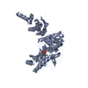
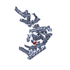
 Assembly
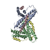

 Mass: 18.015 Da / Num. of mol.: 26 / Source method: isolated from a natural source / Formula: H2O
 Sample preparation
 Processing