 Movie
Movie Controller


+ Open data

- Basic information
| Entry | Database: PDB / ID: 3rfb | ||||||
|---|---|---|---|---|---|---|---|
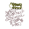
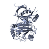
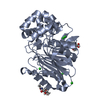
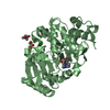
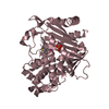
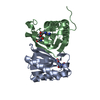
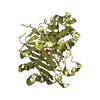
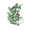
| Title | Structure of fRMsr | ||||||
 Components Components | Putative uncharacterized protein | ||||||
 Keywords Keywords | OXIDOREDUCTASE / fRMsr / GAF / SME | ||||||
| Function / homology |  Function and homology information Function and homology information | ||||||
| Biological species |   Streptococcus pneumoniae (bacteria) Streptococcus pneumoniae (bacteria) | ||||||
| Method |  X-RAY DIFFRACTION / SYNCHROTRON / MOLECULAR REPLACEMENT / Resolution: 2.3 Å X-RAY DIFFRACTION / SYNCHROTRON / MOLECULAR REPLACEMENT / Resolution: 2.3 Å | ||||||
 Authors Authors | Bong, S.M. / Chi, Y.M. | ||||||
 Citation Citation | Journal: to be published Title: Structure of fRMsr Authors: Bong, S.M. / Chi, Y.M. | ||||||
| History |
|
- Structure visualization
| Structure viewer | Molecule: MolmilJmol/JSmol |
|---|
- Downloads & links
-Download
| PDBx/mmCIF format | 3rfb.cif.gz | 76.7 KB | Display | PDBx/mmCIF format |
|---|---|---|---|---|
| PDB format | pdb3rfb.ent.gz | 57.3 KB | Display | PDB format |
| PDBx/mmJSON format | 3rfb.json.gz | Tree view | PDBx/mmJSON format | |
| Others |  Other downloads Other downloads |
-Validation report
| Arichive directory | https://data.pdbj.org/pub/pdb/validation_reports/rf/3rfbftp://data.pdbj.org/pub/pdb/validation_reports/rf/3rfb | HTTPS FTP |
|---|
-Related structure data
| Related structure data |  1vhmS S: Starting model for refinement |
|---|---|
| Similar structure data |


-Links
 PDBj
PDBj

- Assembly
| Deposited unit | 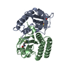
| ||||||||
|---|---|---|---|---|---|---|---|---|---|
| 1 |
| ||||||||
| Unit cell |
|
-Components
| #1: Protein | Mass: 19263.857 Da / Num. of mol.: 2 Source method: isolated from a genetically manipulated source Source: (gene. exp.) Streptococcus pneumoniae (bacteria) / Strain: R6 / Gene: spr0768 / Production host: References: UniProt: Q8DQA6, L-methionine (R)-S-oxide reductase #2: Chemical |   Type: L-peptide linking / Mass: 165.211 Da / Num. of mol.: 2 / Source method: obtained synthetically / Formula: C5H11NO3S Type: L-peptide linking / Mass: 165.211 Da / Num. of mol.: 2 / Source method: obtained synthetically / Formula: C5H11NO3S#3: Water | ChemComp-HOH / |  Mass: 18.015 Da / Num. of mol.: 80 / Source method: isolated from a natural source / Formula: H2O Mass: 18.015 Da / Num. of mol.: 80 / Source method: isolated from a natural source / Formula: H2O |
|---|
-Experimental details
-Experiment
| Experiment | Method: X-RAY DIFFRACTION / Number of used crystals: 1 |
|---|
- Sample preparation
| Crystal | Density Matthews: 2.68 Å3/Da / Density % sol: 54.18 % |
|---|---|
| Crystal grow | Temperature: 295 K / Method: vapor diffusion, hanging drop / pH: 5.5 Details: PEG 400, Ammonium acetate, sodium citrate tribasic dehydrate, pH 5.5, VAPOR DIFFUSION, HANGING DROP, temperature 295K |
-Data collection
| Diffraction source | Source: SYNCHROTRON / Site: PAL/PLS  / Beamline: 6C1 / Wavelength: 1.23968 Å / Beamline: 6C1 / Wavelength: 1.23968 Å |
|---|---|
| Detector | Type: ADSC QUANTUM 210 / Detector: CCD / Date: Oct 13, 2010 |
| Radiation | Protocol: SINGLE WAVELENGTH / Monochromatic (M) / Laue (L): M / Scattering type: x-ray |
| Radiation wavelength | Wavelength: 1.23968 Å / Relative weight: 1 |
| Reflection | Resolution: 2.3→50 Å / Num. all: 18660 / Num. obs: 18660 / % possible obs: 99.6 % / Observed criterion σ(F): 0 / Observed criterion σ(I): 0 |
| Reflection shell | Resolution: 2.3→2.44 Å / % possible all: 99.5 |
- Processing
| Software |
| ||||||||||||||||||||
|---|---|---|---|---|---|---|---|---|---|---|---|---|---|---|---|---|---|---|---|---|---|
| Refinement | Method to determine structure: MOLECULAR REPLACEMENT Starting model: 1VHM Resolution: 2.3→37.14 Å / σ(F): 0 / Stereochemistry target values: Engh & Huber
| ||||||||||||||||||||
| Refinement step | Cycle: LAST / Resolution: 2.3→37.14 Å
| ||||||||||||||||||||
| Refine LS restraints |
|
