 Movie
Movie Controller

[English] 日本語
 Yorodumi
- PDB-3rdy: Crystal Structure of buckwheat trypsin inhibitor rBTI at 1.84 ang... -
+ Open data

- Basic information
| Entry | Database: PDB / ID: 3rdy | ||||||
|---|---|---|---|---|---|---|---|
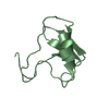
| Title | Crystal Structure of buckwheat trypsin inhibitor rBTI at 1.84 angstrom resolution | ||||||
 Components Components | BWI-1=PROTEASE inhibitor/trypsin inhibitor | ||||||
 Keywords Keywords | HYDROLASE INHIBITOR / serine protease inhibitor / potato inhibitor I / trypsin inhibitor / trypsin | ||||||
| Function / homology |  Function and homology information Function and homology information | ||||||
| Biological species |  Fagopyrum esculentum (common buckwheat) Fagopyrum esculentum (common buckwheat) | ||||||
| Method |  X-RAY DIFFRACTION / SYNCHROTRON / MOLECULAR REPLACEMENT / Resolution: 1.84 Å X-RAY DIFFRACTION / SYNCHROTRON / MOLECULAR REPLACEMENT / Resolution: 1.84 Å | ||||||
 Authors Authors | Wang, L.F. / Li, M. / Chang, W.R. | ||||||
 Citation Citation | Journal: Plos One / Year: 2011 Title: Conformational Changes of rBTI from Buckwheat upon Binding to Trypsin: Implications for the Role of the P(8)' Residue in the Potato Inhibitor I Family Authors: Wang, L.F. / Zhao, F. / Li, M. / Zhang, H. / Gao, Y. / Cao, P. / Pan, X. / Wang, Z. / Chang, W.R. | ||||||
| History |
|
- Structure visualization
| Structure viewer | Molecule: MolmilJmol/JSmol |
|---|
- Downloads & links
-Download
| PDBx/mmCIF format | 3rdy.cif.gz | 27.5 KB | Display | PDBx/mmCIF format |
|---|---|---|---|---|
| PDB format | pdb3rdy.ent.gz | 16.7 KB | Display | PDB format |
| PDBx/mmJSON format | 3rdy.json.gz | Tree view | PDBx/mmJSON format | |
| Others |  Other downloads Other downloads |
-Validation report
| Arichive directory | https://data.pdbj.org/pub/pdb/validation_reports/rd/3rdyftp://data.pdbj.org/pub/pdb/validation_reports/rd/3rdy | HTTPS FTP |
|---|
-Related structure data
| Related structure data |  3rdzSC S: Starting model for refinement C: citing same article ( |
|---|---|
| Similar structure data |


-Links
 PDBj
PDBj
- Assembly
| Deposited unit | 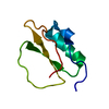
| ||||||||
|---|---|---|---|---|---|---|---|---|---|
| 1 |
| ||||||||
| Unit cell |
|
-Components
| #1: Protein | Mass: 9019.240 Da / Num. of mol.: 1 Source method: isolated from a genetically manipulated source Source: (gene. exp.) Fagopyrum esculentum (common buckwheat)Gene: BWI-1, Edn1 / Plasmid: QIA express pQE-31 / Production host:  |
|---|---|
| #2: Water | ChemComp-HOH /  Mass: 18.015 Da / Num. of mol.: 92 / Source method: isolated from a natural source / Formula: H2O Mass: 18.015 Da / Num. of mol.: 92 / Source method: isolated from a natural source / Formula: H2O |
| Has protein modification | Y |
-Experimental details
-Experiment
| Experiment | Method: X-RAY DIFFRACTION / Number of used crystals: 1 |
|---|
- Sample preparation
| Crystal | Density Matthews: 2.5 Å3/Da / Density % sol: 50.75 % |
|---|---|
| Crystal grow | Temperature: 291 K / Method: vapor diffusion, hanging drop / pH: 4.4 Details: 24% (w/v) PEG MME 2000, 220mM ammonium sulfate, 100mM sodium acetate (pH 4.4), 100mM sodium iodide, VAPOR DIFFUSION, HANGING DROP, temperature 291K |
-Data collection
| Diffraction | Mean temperature: 100 K |
|---|---|
| Diffraction source | Source: SYNCHROTRON / Site: BSRF  / Beamline: 1W2B / Wavelength: 1 Å / Beamline: 1W2B / Wavelength: 1 Å |
| Detector | Type: MAR555 FLAT PANEL / Detector: IMAGE PLATE / Date: Dec 3, 2009 |
| Radiation | Monochromator: Si(111) double flat-crystal monochromator / Protocol: SINGLE WAVELENGTH / Monochromatic (M) / Laue (L): M / Scattering type: x-ray |
| Radiation wavelength | Wavelength: 1 Å / Relative weight: 1 |
| Reflection | Resolution: 1.84→15 Å / Num. all: 8360 / Num. obs: 8344 / % possible obs: 99.8 % / Observed criterion σ(F): 2 / Observed criterion σ(I): 2 / Redundancy: 24.1 % / Biso Wilson estimate: 24.4 Å2 / Rmerge(I) obs: 0.08 / Net I/σ(I): 46.4 |
| Reflection shell | Resolution: 1.84→1.91 Å / Redundancy: 23.2 % / Rmerge(I) obs: 0.5 / Mean I/σ(I) obs: 4.7 / % possible all: 100 |
- Processing
| Software |
| ||||||||||||||||||||||||||||
|---|---|---|---|---|---|---|---|---|---|---|---|---|---|---|---|---|---|---|---|---|---|---|---|---|---|---|---|---|---|
| Refinement | Method to determine structure: MOLECULAR REPLACEMENT Starting model: PDB ENTRY 3RDZ Resolution: 1.84→14.464 Å / FOM work R set: 0.856 / SU ML: 0.16 / Isotropic thermal model: isotropic / Cross valid method: THROUGHOUT / σ(F): 2 / Phase error: 20.01 / Stereochemistry target values: ML
| ||||||||||||||||||||||||||||
| Solvent computation | Shrinkage radii: 0.9 Å / VDW probe radii: 1.11 Å / Solvent model: FLAT BULK SOLVENT MODEL / Bsol: 64.987 Å2 / ksol: 0.393 e/Å3 | ||||||||||||||||||||||||||||
| Displacement parameters | Biso mean: 28.8 Å2
| ||||||||||||||||||||||||||||
| Refinement step | Cycle: LAST / Resolution: 1.84→14.464 Å
| ||||||||||||||||||||||||||||
| Refine LS restraints |
| ||||||||||||||||||||||||||||
| LS refinement shell |
|
