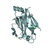
- PDB-3rbq: Co-crystal structure of human UNC119 (retina gene 4) and an N-ter... -
+
Open data
ID or keywords:
Loading...
-
Basic information
Entry
Database: PDB / ID: 3rbq
Title
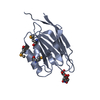
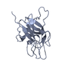
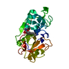
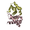
Co-crystal structure of human UNC119 (retina gene 4) and an N-terminal Transducin-alpha mimicking peptide
Components
Guanine nucleotide-binding protein G(t) subunit alpha-1
Protein unc-119 homolog A
Keywords
Protein Transport/Lipid Binding Protein / UNC(uncoordinated)119 homolog C. elegans / G protein trafficking / acyl binnding protein / light-induced translocation / UNC119 deletion / Protein Transport-Lipid Binding Protein complex
Function / homology
Function and homology information
background adaptation / neural tissue regeneration / negative regulation of caveolin-mediated endocytosis / negative regulation of clathrin-dependent endocytosis / negative regulation of cyclic-nucleotide phosphodiesterase activity / retinal rod cell differentiation / retinal cone cell differentiation / visual behavior / regulation of opsin-mediated signaling pathway / cellular response to electrical stimulus ...background adaptation / neural tissue regeneration / negative regulation of caveolin-mediated endocytosis / negative regulation of clathrin-dependent endocytosis / negative regulation of cyclic-nucleotide phosphodiesterase activity / retinal rod cell differentiation / retinal cone cell differentiation / visual behavior / regulation of opsin-mediated signaling pathway / cellular response to electrical stimulus / photoreceptor connecting cilium / G protein-coupled opsin signaling pathway / sensory perception of umami taste / detection of light stimulus involved in visual perception / eye photoreceptor cell development / dopamine secretion / detection of chemical stimulus involved in sensory perception of bitter taste / lipoprotein transport / photoreceptor outer segment membrane / positive regulation of protein tyrosine kinase activity / spindle midzone / acyl binding / mitotic cytokinesis / response to light stimulus / phototransduction, visible light / phototransduction / photoreceptor outer segment / intercellular bridge / photoreceptor inner segment / visual perception / G protein-coupled receptor binding / adenylate cyclase-modulating G protein-coupled receptor signaling pathway / G-protein beta/gamma-subunit complex binding / Activation of the phototransduction cascade / endocytosis / spindle pole / photoreceptor disc membrane / GDP binding / Inactivation, recovery and regulation of the phototransduction cascade / nervous system development / heterotrimeric G-protein complex / G alpha (i) signalling events / chemical synaptic transmission / cell population proliferation / apical plasma membrane / neuronal cell body / GTPase activity / lipid binding / synapse / centrosome / protein kinase binding / GTP binding / signal transduction / metal ion binding / membrane / plasma membrane / cytosol / cytoplasm Similarity search - Function
: / GMP phosphodiesterase, delta subunit / GMP phosphodiesterase, delta subunit / GMP phosphodiesterase, delta subunit superfamily / GMP-PDE, delta subunit / Coagulation Factor XIII; Chain A, domain 1 / G-protein alpha subunit, group I / Distorted Sandwich / Guanine nucleotide binding protein (G-protein), alpha subunit / G protein alpha subunit, helical insertion ...: / GMP phosphodiesterase, delta subunit / GMP phosphodiesterase, delta subunit / GMP phosphodiesterase, delta subunit superfamily / GMP-PDE, delta subunit / Coagulation Factor XIII; Chain A, domain 1 / G-protein alpha subunit, group I / Distorted Sandwich / Guanine nucleotide binding protein (G-protein), alpha subunit / G protein alpha subunit, helical insertion / G-protein alpha subunit / G-alpha domain profile. / G protein alpha subunit / Immunoglobulin E-set / P-loop containing nucleoside triphosphate hydrolase / Mainly Beta Similarity search - Domain/homology
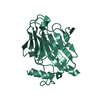
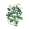
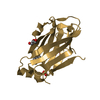
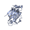
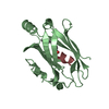
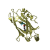
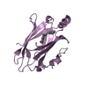
A: Protein unc-119 homolog A B: Protein unc-119 homolog A C: Protein unc-119 homolog A D: Protein unc-119 homolog A E: Protein unc-119 homolog A F: Protein unc-119 homolog A G: Guanine nucleotide-binding protein G(t) subunit alpha-1 H: Guanine nucleotide-binding protein G(t) subunit alpha-1 I: Guanine nucleotide-binding protein G(t) subunit alpha-1 J: Guanine nucleotide-binding protein G(t) subunit alpha-1 K: Guanine nucleotide-binding protein G(t) subunit alpha-1 L: Guanine nucleotide-binding protein G(t) subunit alpha-1
Resolution: 2→29.37 Å / Cor.coef. Fo:Fc: 0.954 / Cor.coef. Fo:Fc free: 0.924 / SU B: 4.036 / SU ML: 0.115 / Cross valid method: THROUGHOUT / ESU R: 0.186 / ESU R Free: 0.174 / Stereochemistry target values: MAXIMUM LIKELIHOOD / Details: HYDROGENS HAVE BEEN ADDED IN THE RIDING POSITIONS
Rfactor
Num. reflection
% reflection
Selection details
Rfree
0.24557
4087
5 %
RANDOM
Rwork
0.18845
-
-
-
obs
0.19134
77030
99.96 %
-
all
-
78089
-
-
Solvent computation
Ion probe radii: 0.8 Å / Shrinkage radii: 0.8 Å / VDW probe radii: 1.4 Å / Solvent model: MASK
Displacement parameters
Biso mean: 30.833 Å2
Baniso -1
Baniso -2
Baniso -3
1-
0.24 Å2
0 Å2
0 Å2
2-
-
0.7 Å2
0 Å2
3-
-
-
-0.94 Å2
Refinement step
Cycle: LAST / Resolution: 2→29.37 Å
Protein
Nucleic acid
Ligand
Solvent
Total
Num. atoms
8494
0
0
793
9287
Refine LS restraints
Refine-ID
Type
Dev ideal
Dev ideal target
Number
X-RAY DIFFRACTION
r_bond_refined_d
0.023
0.022
8797
X-RAY DIFFRACTION
r_bond_other_d
X-RAY DIFFRACTION
r_angle_refined_deg
1.843
1.955
11834
X-RAY DIFFRACTION
r_angle_other_deg
X-RAY DIFFRACTION
r_dihedral_angle_1_deg
6.252
5
1023
X-RAY DIFFRACTION
r_dihedral_angle_2_deg
34.778
22.966
472
X-RAY DIFFRACTION
r_dihedral_angle_3_deg
14.773
15
1449
X-RAY DIFFRACTION
r_dihedral_angle_4_deg
17.78
15
76
X-RAY DIFFRACTION
r_chiral_restr
0.15
0.2
1204
X-RAY DIFFRACTION
r_gen_planes_refined
0.011
0.021
6860
X-RAY DIFFRACTION
r_gen_planes_other
X-RAY DIFFRACTION
r_nbd_refined
X-RAY DIFFRACTION
r_nbd_other
X-RAY DIFFRACTION
r_nbtor_refined
X-RAY DIFFRACTION
r_nbtor_other
X-RAY DIFFRACTION
r_xyhbond_nbd_refined
X-RAY DIFFRACTION
r_xyhbond_nbd_other
X-RAY DIFFRACTION
r_metal_ion_refined
X-RAY DIFFRACTION
r_metal_ion_other
X-RAY DIFFRACTION
r_symmetry_vdw_refined
X-RAY DIFFRACTION
r_symmetry_vdw_other
X-RAY DIFFRACTION
r_symmetry_hbond_refined
X-RAY DIFFRACTION
r_symmetry_hbond_other
X-RAY DIFFRACTION
r_symmetry_metal_ion_refined
X-RAY DIFFRACTION
r_symmetry_metal_ion_other
X-RAY DIFFRACTION
r_mcbond_it
1.339
1.5
5155
X-RAY DIFFRACTION
r_mcbond_other
X-RAY DIFFRACTION
r_mcangle_it
2.322
2
8355
X-RAY DIFFRACTION
r_scbond_it
3.452
3
3642
X-RAY DIFFRACTION
r_scangle_it
5.441
4.5
3479
X-RAY DIFFRACTION
r_rigid_bond_restr
X-RAY DIFFRACTION
r_sphericity_free
X-RAY DIFFRACTION
r_sphericity_bonded
LS refinement shell
Resolution: 2→2.052 Å / Total num. of bins used: 20
Rfactor
Num. reflection
% reflection
Rfree
0.28
288
-
Rwork
0.213
5583
-
obs
-
-
99.97 %
+
About Yorodumi
-
News
-
Feb 9, 2022. New format data for meta-information of EMDB entries
New format data for meta-information of EMDB entries
Version 3 of the EMDB header file is now the official format.
The previous official version 1.9 will be removed from the archive.
In the structure databanks used in Yorodumi, some data are registered as the other names, "COVID-19 virus" and "2019-nCoV". Here are the details of the virus and the list of structure data.
Jan 31, 2019. EMDB accession codes are about to change! (news from PDBe EMDB page)
EMDB accession codes are about to change! (news from PDBe EMDB page)
The allocation of 4 digits for EMDB accession codes will soon come to an end. Whilst these codes will remain in use, new EMDB accession codes will include an additional digit and will expand incrementally as the available range of codes is exhausted. The current 4-digit format prefixed with “EMD-” (i.e. EMD-XXXX) will advance to a 5-digit format (i.e. EMD-XXXXX), and so on. It is currently estimated that the 4-digit codes will be depleted around Spring 2019, at which point the 5-digit format will come into force.
The EM Navigator/Yorodumi systems omit the EMD- prefix.
Related info.:Q: What is EMD? / ID/Accession-code notation in Yorodumi/EM Navigator
Yorodumi is a browser for structure data from EMDB, PDB, SASBDB, etc.
This page is also the successor to EM Navigator detail page, and also detail information page/front-end page for Omokage search.
The word "yorodu" (or yorozu) is an old Japanese word meaning "ten thousand". "mi" (miru) is to see.
Related info.:EMDB / PDB / SASBDB / Comparison of 3 databanks / Yorodumi Search / Aug 31, 2016. New EM Navigator & Yorodumi / Yorodumi Papers / Jmol/JSmol / Function and homology information / Changes in new EM Navigator and Yorodumi
 Movie
Movie Controller

 Yorodumi
 Open data
 Basic information
 Components
 Keywords
 Function and homology information
 Homo sapiens (human)
 X-RAY DIFFRACTION /
  Authors
 Citation
 Structure visualization
 Downloads & links
 Other downloads
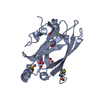
 PDBj
PDBj


 Assembly
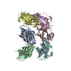


 Mass: 18.015 Da / Num. of mol.: 793 / Source method: isolated from a natural source / Formula: H2O
 Sample preparation
 / Beamline: BL9-2 / Wavelength: 0.9749 Å
 Processing