 Movie
Movie Controller

[English] 日本語
 Yorodumi
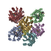
- PDB-3qs7: Crystal structure of a human Flt3 ligand-receptor ternary complex -
+ Open data

- Basic information
| Entry | Database: PDB / ID: 3qs7 | ||||||
|---|---|---|---|---|---|---|---|
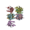
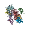
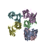
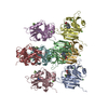
| Title | Crystal structure of a human Flt3 ligand-receptor ternary complex | ||||||
 Components Components |
| ||||||
 Keywords Keywords | CYTOKINE/SIGNALING PROTEIN / immunoglobulin-like domain / four-helical bundle cytokine / cytokine-receptor complex / extracellular complex / receptor tyrosine kinase / CYTOKINE-SIGNALING PROTEIN complex | ||||||
| Function / homology |  Function and homology information Function and homology informationFLT3 mutants bind TKIs / KW2449-resistant FLT3 mutants / semaxanib-resistant FLT3 mutants / crenolanib-resistant FLT3 mutants / gilteritinib-resistant FLT3 mutants / lestaurtinib-resistant FLT3 mutants / midostaurin-resistant FLT3 mutants / pexidartinib-resistant FLT3 mutants / ponatinib-resistant FLT3 mutants / quizartinib-resistant FLT3 mutants ...FLT3 mutants bind TKIs / KW2449-resistant FLT3 mutants / semaxanib-resistant FLT3 mutants / crenolanib-resistant FLT3 mutants / gilteritinib-resistant FLT3 mutants / lestaurtinib-resistant FLT3 mutants / midostaurin-resistant FLT3 mutants / pexidartinib-resistant FLT3 mutants / ponatinib-resistant FLT3 mutants / quizartinib-resistant FLT3 mutants / sorafenib-resistant FLT3 mutants / sunitinib-resistant FLT3 mutants / tandutinib-resistant FLT3 mutants / linifanib-resistant FLT3 mutants / tamatinib-resistant FLT3 mutants / leukocyte homeostasis / lymphocyte proliferation / common myeloid progenitor cell proliferation / pro-B cell differentiation / vascular endothelial growth factor receptor activity / dendritic cell differentiation / nuclear glucocorticoid receptor binding / STAT5 Activation / phosphatidylinositol 3-kinase activator activity / myeloid progenitor cell differentiation / FLT3 signaling through SRC family kinases / cytokine receptor activity / embryonic hemopoiesis / positive regulation of tyrosine phosphorylation of STAT protein / cellular response to glucocorticoid stimulus / STAT5 activation downstream of FLT3 ITD mutants / growth factor binding / cellular response to cytokine stimulus / positive regulation of MAP kinase activity / positive regulation of natural killer cell proliferation / hemopoiesis / PI3K Cascade / Signaling by FLT3 ITD and TKD mutants / peptidyl-tyrosine phosphorylation / transmembrane receptor protein tyrosine kinase activity / FLT3 Signaling / FLT3 signaling by CBL mutants / liver regeneration / Negative regulation of FLT3 / cell surface receptor protein tyrosine kinase signaling pathway / B cell differentiation / cytokine activity / receptor protein-tyrosine kinase / receptor tyrosine kinase binding / Constitutive Signaling by Aberrant PI3K in Cancer / cytokine-mediated signaling pathway / protein autophosphorylation / PIP3 activates AKT signaling / cell migration / PI5P, PP2A and IER3 Regulate PI3K/AKT Signaling / RAF/MAP kinase cascade / protein tyrosine kinase activity / regulation of apoptotic process / positive regulation of MAPK cascade / positive regulation of phosphatidylinositol 3-kinase/protein kinase B signal transduction / signaling receptor complex / endosome membrane / endoplasmic reticulum lumen / signaling receptor binding / positive regulation of cell population proliferation / protein-containing complex binding / cell surface / signal transduction / endoplasmic reticulum / : / extracellular region / ATP binding / membrane / plasma membrane / cytosol Similarity search - Function | ||||||
| Biological species |  Homo sapiens (human) Homo sapiens (human) | ||||||
| Method |  X-RAY DIFFRACTION / SYNCHROTRON / MOLECULAR REPLACEMENT / molecular replacement / Resolution: 4.3 Å X-RAY DIFFRACTION / SYNCHROTRON / MOLECULAR REPLACEMENT / molecular replacement / Resolution: 4.3 Å | ||||||
 Authors Authors | Verstraete, K. / Savvides, S.N. | ||||||
 Citation Citation | Journal: Blood / Year: 2011 Title: Structural insights into the extracellular assembly of the hematopoietic Flt3 signaling complex. Authors: Verstraete, K. / Vandriessche, G. / Januar, M. / Elegheert, J. / Shkumatov, A.V. / Desfosses, A. / Van Craenenbroeck, K. / Svergun, D.I. / Gutsche, I. / Vergauwen, B. / Savvides, S.N. #1: Journal: To be PublishedTitle: Inducible production of human Flt3 ectodomain variants in mammalian cells and preliminary crystallographic analysis of Flt3 ligand-receptor complexes Authors: Verstraete, K. / Vandriessche, G. / Januar, M. / Elegheert, J. / Shkumatov, A. / Desfosses, A. / Van Craenenbroeck, K. / Svergun, D. / Gutsche, I. / Vergauwen, B. / Savvides, S.N. #2: Journal: Protein J. / Year: 2009 Title: Efficient production of bioactive recombinant human Flt3 ligand in E. coli. Authors: Verstraete, K. / Koch, S. / Ertugrul, S. / Vandenberghe, I. / Aerts, M. / Vandriessche, G. / Thiede, C. / Savvides, S.N. | ||||||
| History |
|
- Structure visualization
| Structure viewer | Molecule: MolmilJmol/JSmol |
|---|
- Downloads & links
-Download
| PDBx/mmCIF format | 3qs7.cif.gz | 283.1 KB | Display | PDBx/mmCIF format |
|---|---|---|---|---|
| PDB format | pdb3qs7.ent.gz | 211.4 KB | Display | PDB format |
| PDBx/mmJSON format | 3qs7.json.gz | Tree view | PDBx/mmJSON format | |
| Others |  Other downloads Other downloads |
-Validation report
| Arichive directory | https://data.pdbj.org/pub/pdb/validation_reports/qs/3qs7ftp://data.pdbj.org/pub/pdb/validation_reports/qs/3qs7 | HTTPS FTP |
|---|
-Related structure data
| Related structure data |  3qs9C  1eteS  2e9wS C: citing same article ( S: Starting model for refinement |
|---|---|
| Similar structure data |


-Links
 PDBj
PDBj


- Assembly
| Deposited unit | 
| ||||||||
|---|---|---|---|---|---|---|---|---|---|
| 1 | 
| ||||||||
| 2 | 
| ||||||||
| Unit cell |
|
-Components
| #1: Protein | Mass: 15873.217 Da / Num. of mol.: 4 / Fragment: extracellular domain (UNP residues 27-160) Source method: isolated from a genetically manipulated source Source: (gene. exp.) Homo sapiens (human) / Gene: FLT3LG / Plasmid: pET15B / Production host:  #2: Protein | Mass: 48099.188 Da / Num. of mol.: 4 / Fragment: extracellular domain (UNP residues 27-436) Source method: isolated from a genetically manipulated source Source: (gene. exp.) Homo sapiens (human) / Gene: FLT3, STK1 / Plasmid: pcDNA4/TO / Cell line (production host): HEK 293 GNTI- / Production host: Homo sapiens (human)References: UniProt: P36888, receptor protein-tyrosine kinase #3: Sugar | ChemComp-NAG /   Type: D-saccharide, beta linking / Mass: 221.208 Da / Num. of mol.: 7 Type: D-saccharide, beta linking / Mass: 221.208 Da / Num. of mol.: 7Source method: isolated from a genetically manipulated source Formula: C8H15NO6 Has protein modification | Y | |
|---|
-Experimental details
-Experiment
| Experiment | Method: X-RAY DIFFRACTION / Number of used crystals: 1 |
|---|
- Sample preparation
| Crystal | Density Matthews: 2.96 Å3/Da / Density % sol: 58.48 % |
|---|---|
| Crystal grow | Temperature: 293 K / Method: vapor diffusion, sitting drop / pH: 6 Details: 11-13% PEG4000, 0.1 M magnesium chloride, 50 mM MES, 5 mg/mL Flt3 ligand-receptor complex, pH 6.0, VAPOR DIFFUSION, SITTING DROP, temperature 293.0K |
-Data collection
| Diffraction | Mean temperature: 100 K |
|---|---|
| Diffraction source | Source: SYNCHROTRON / Site: ESRF  / Beamline: ID23-1 / Wavelength: 0.9714 / Beamline: ID23-1 / Wavelength: 0.9714 |
| Detector | Type: ADSC QUANTUM 315r / Detector: CCD / Date: Mar 12, 2010 |
| Radiation | Protocol: SINGLE WAVELENGTH / Monochromatic (M) / Laue (L): M / Scattering type: x-ray |
| Radiation wavelength | Wavelength: 0.9714 Å / Relative weight: 1 |
| Reflection | Resolution: 4.3→40 Å / Num. all: 20434 / Num. obs: 20184 / % possible obs: 98.8 % / Observed criterion σ(I): -3 / Redundancy: 3.8 % / Biso Wilson estimate: 135 Å2 / Rmerge(I) obs: 0.108 / Net I/σ(I): 12.04 |
| Reflection shell | Resolution: 4.3→4.45 Å / Redundancy: 3.8 % / Rmerge(I) obs: 0.759 / Mean I/σ(I) obs: 2.08 / % possible all: 98.9 |
-Phasing
| Phasing | Method: molecular replacement |
|---|
- Processing
| Software |
| ||||||||||||||||||||||||||||||||||||||||||||||||||||||||||||||||||||||||
|---|---|---|---|---|---|---|---|---|---|---|---|---|---|---|---|---|---|---|---|---|---|---|---|---|---|---|---|---|---|---|---|---|---|---|---|---|---|---|---|---|---|---|---|---|---|---|---|---|---|---|---|---|---|---|---|---|---|---|---|---|---|---|---|---|---|---|---|---|---|---|---|---|---|
| Refinement | Method to determine structure: MOLECULAR REPLACEMENT Starting model: PDB ENTRIES 1ETE AND 2E9W Resolution: 4.3→39.04 Å / Cor.coef. Fo:Fc: 0.8832 / Cor.coef. Fo:Fc free: 0.8516 / Occupancy max: 1 / Occupancy min: 1 / Isotropic thermal model: ISOTROPIC / Cross valid method: THROUGHOUT / σ(F): 0 / Stereochemistry target values: Engh & Huber
| ||||||||||||||||||||||||||||||||||||||||||||||||||||||||||||||||||||||||
| Displacement parameters | Biso max: 242.95 Å2 / Biso mean: 168.8768 Å2 / Biso min: 122.08 Å2
| ||||||||||||||||||||||||||||||||||||||||||||||||||||||||||||||||||||||||
| Refine analyze | Luzzati coordinate error obs: 1.374 Å | ||||||||||||||||||||||||||||||||||||||||||||||||||||||||||||||||||||||||
| Refinement step | Cycle: LAST / Resolution: 4.3→39.04 Å
| ||||||||||||||||||||||||||||||||||||||||||||||||||||||||||||||||||||||||
| Refine LS restraints |
| ||||||||||||||||||||||||||||||||||||||||||||||||||||||||||||||||||||||||
| LS refinement shell | Resolution: 4.3→4.53 Å / Total num. of bins used: 10
|
