 Movie
Movie Controller


+ Open data

- Basic information
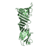
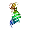
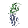
| Entry | Database: PDB / ID: 1ete | ||||||
|---|---|---|---|---|---|---|---|
| Title | CRYSTAL STRUCTURE OF THE FLT3 LIGAND | ||||||
 Components Components | FLT3 LIGAND | ||||||
 Keywords Keywords | CYTOKINE / four-helix bundle | ||||||
| Function / homology |  Function and homology information Function and homology informationdendritic cell differentiation / STAT5 Activation / FLT3 signaling through SRC family kinases / embryonic hemopoiesis / positive regulation of natural killer cell proliferation / PI3K Cascade / FLT3 Signaling / FLT3 signaling by CBL mutants / Negative regulation of FLT3 / B cell differentiation ...dendritic cell differentiation / STAT5 Activation / FLT3 signaling through SRC family kinases / embryonic hemopoiesis / positive regulation of natural killer cell proliferation / PI3K Cascade / FLT3 Signaling / FLT3 signaling by CBL mutants / Negative regulation of FLT3 / B cell differentiation / cytokine activity / receptor tyrosine kinase binding / Constitutive Signaling by Aberrant PI3K in Cancer / PIP3 activates AKT signaling / PI5P, PP2A and IER3 Regulate PI3K/AKT Signaling / RAF/MAP kinase cascade / signaling receptor binding / positive regulation of cell population proliferation / cell surface / signal transduction / : / extracellular region / membrane / plasma membrane / cytosol Similarity search - Function | ||||||
| Biological species |  Homo sapiens (human) Homo sapiens (human) | ||||||
| Method |  X-RAY DIFFRACTION / SYNCHROTRON / Resolution: 2.2 Å X-RAY DIFFRACTION / SYNCHROTRON / Resolution: 2.2 Å | ||||||
 Authors Authors | Savvides, S.N. / Boone, T. / Karplus, P.A. | ||||||
 Citation Citation | Journal: Nat.Struct.Biol. / Year: 2000 Title: Flt3 ligand structure and unexpected commonalities of helical bundles and cystine knots. Authors: Savvides, S.N. / Boone, T. / Andrew Karplus, P. | ||||||
| History |
|
- Structure visualization
| Structure viewer | Molecule: MolmilJmol/JSmol |
|---|
- Downloads & links
-Download
| PDBx/mmCIF format | 1ete.cif.gz | 121.2 KB | Display | PDBx/mmCIF format |
|---|---|---|---|---|
| PDB format | pdb1ete.ent.gz | 95.8 KB | Display | PDB format |
| PDBx/mmJSON format | 1ete.json.gz | Tree view | PDBx/mmJSON format | |
| Others |  Other downloads Other downloads |
-Validation report
| Arichive directory | https://data.pdbj.org/pub/pdb/validation_reports/et/1eteftp://data.pdbj.org/pub/pdb/validation_reports/et/1ete | HTTPS FTP |
|---|
-Related structure data
| Similar structure data |
|---|


-Links
 PDBj
PDBj


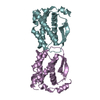
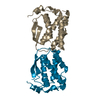
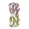
- Assembly
| Deposited unit | 
| ||||||||
|---|---|---|---|---|---|---|---|---|---|
| 1 | 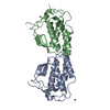
| ||||||||
| 2 | 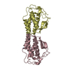
| ||||||||
| 3 | 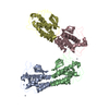
| ||||||||
| 4 | 
| ||||||||
| Unit cell |
|
-Components
| #1: Protein | Mass: 15445.717 Da / Num. of mol.: 4 / Fragment: RECEPTOR BINDING DOMAIN Source method: isolated from a genetically manipulated source Source: (gene. exp.) Homo sapiens (human) / Production host: Bacteria (eubacteria) / References: UniProt: P49771#2: Chemical | ChemComp-ZN /   Mass: 65.409 Da / Num. of mol.: 9 / Source method: obtained synthetically / Formula: Zn Mass: 65.409 Da / Num. of mol.: 9 / Source method: obtained synthetically / Formula: Zn#3: Water | ChemComp-HOH / |  Mass: 18.015 Da / Num. of mol.: 214 / Source method: isolated from a natural source / Formula: H2O Mass: 18.015 Da / Num. of mol.: 214 / Source method: isolated from a natural source / Formula: H2OHas protein modification | Y | Nonpolymer details | WATER 6049 WAS MODELED IN WHAT IS BELIEVED TO BE A LOW OCCUPANCY SITE FOR AN ORDERED ZINC ATOM. | |
|---|
-Experimental details
-Experiment
| Experiment | Method: X-RAY DIFFRACTION / Number of used crystals: 1 |
|---|
- Sample preparation
| Crystal | Density Matthews: 2.32 Å3/Da / Density % sol: 46.98 % | |||||||||||||||||||||||||||||||||||
|---|---|---|---|---|---|---|---|---|---|---|---|---|---|---|---|---|---|---|---|---|---|---|---|---|---|---|---|---|---|---|---|---|---|---|---|---|
| Crystal grow | Temperature: 298 K / Method: vapor diffusion, hanging drop / pH: 6.4 Details: PEG 8000, zinc acetate, sodium cacodylate, pH 6.4, VAPOR DIFFUSION, HANGING DROP, temperature 298.0K | |||||||||||||||||||||||||||||||||||
| Crystal grow | *PLUS Method: vapor diffusionDetails: drop consists of 1:1 mixture of well and protein solutions | |||||||||||||||||||||||||||||||||||
| Components of the solutions | *PLUS
|
-Data collection
| Diffraction | Mean temperature: 100 K |
|---|---|
| Diffraction source | Source: SYNCHROTRON / Site: CHESS  / Beamline: F1 / Wavelength: 0.92 / Beamline: F1 / Wavelength: 0.92 |
| Detector | Type: PRINCETON 2K / Detector: CCD / Date: Jan 25, 1996 |
| Radiation | Protocol: SINGLE WAVELENGTH / Monochromatic (M) / Laue (L): M / Scattering type: x-ray |
| Radiation wavelength | Wavelength: 0.92 Å / Relative weight: 1 |
| Reflection | Resolution: 2.2→20 Å / Num. all: 117033 / Num. obs: 29421 / % possible obs: 96.9 % / Observed criterion σ(I): 3.8 / Redundancy: 4 % / Rmerge(I) obs: 0.062 / Net I/σ(I): 12 |
| Reflection shell | Resolution: 2.2→2.24 Å / Redundancy: 3.3 % / Rmerge(I) obs: 0.221 / Num. unique all: 1372 / % possible all: 95.5 |
| Reflection shell | *PLUS % possible obs: 95.5 % / Mean I/σ(I) obs: 3.8 |
- Processing
| Software |
| ||||||||||||||||||||
|---|---|---|---|---|---|---|---|---|---|---|---|---|---|---|---|---|---|---|---|---|---|
| Refinement | Resolution: 2.2→20 Å / σ(F): 1 / σ(I): 0 / Stereochemistry target values: Engh & Huber
| ||||||||||||||||||||
| Refinement step | Cycle: LAST / Resolution: 2.2→20 Å
| ||||||||||||||||||||
| Refine LS restraints |
|
