 Movie
Movie Controller

[English] 日本語
 Yorodumi
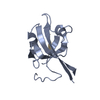
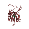
- PDB-3qjn: Structural flexibility of Shank PDZ domain is important for its b... -
+ Open data

- Basic information
| Entry | Database: PDB / ID: 3qjn | ||||||
|---|---|---|---|---|---|---|---|
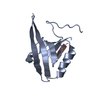
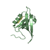
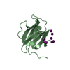
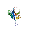
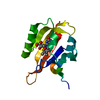
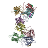
| Title | Structural flexibility of Shank PDZ domain is important for its binding to different ligands | ||||||
 Components Components |
| ||||||
 Keywords Keywords | PROTEIN BINDING / PDZ domain / Protein-protein interaction / Beta-PIX | ||||||
| Function / homology |  Function and homology information Function and homology informationsomatostatin receptor binding / determination of affect / negative regulation of microtubule nucleation / Ephrin signaling / presynaptic actin cytoskeleton organization / EGFR downregulation / RHOU GTPase cycle / RHOV GTPase cycle / NRAGE signals death through JNK / G alpha (12/13) signalling events ...somatostatin receptor binding / determination of affect / negative regulation of microtubule nucleation / Ephrin signaling / presynaptic actin cytoskeleton organization / EGFR downregulation / RHOU GTPase cycle / RHOV GTPase cycle / NRAGE signals death through JNK / G alpha (12/13) signalling events / RHOQ GTPase cycle / synaptic receptor adaptor activity / RAC1 GTPase cycle / olfactory behavior / postsynaptic actin cytoskeleton organization / RHOA GTPase cycle / synapse maturation / Neurexins and neuroligins / storage vacuole / astrocyte cell migration / structural constituent of postsynaptic density / negative regulation of actin filament bundle assembly / positive regulation of growth hormone secretion / righting reflex / habituation / protein localization to synapse / dendritic spine morphogenesis / ankyrin repeat binding / vocalization behavior / gamma-tubulin binding / ATP-dependent protein binding / AMPA selective glutamate receptor signaling pathway / lamellipodium assembly / neuromuscular process controlling balance / small GTPase-mediated signal transduction / mitotic spindle pole / Golgi organization / positive regulation of dendritic spine development / adult behavior / associative learning / social behavior / hematopoietic progenitor cell differentiation / long-term memory / Rho protein signal transduction / ruffle / excitatory synapse / guanyl-nucleotide exchange factor activity / ionotropic glutamate receptor binding / positive regulation of excitatory postsynaptic potential / SH3 domain binding / synapse organization / G protein-coupled receptor binding / modulation of chemical synaptic transmission / Schaffer collateral - CA1 synapse / GABA-ergic synapse / lamellipodium / growth cone / signaling receptor complex adaptor activity / protein-containing complex assembly / scaffold protein binding / cell cortex / dendritic spine / postsynaptic membrane / neuron projection / postsynaptic density / postsynapse / positive regulation of apoptotic process / focal adhesion / neuronal cell body / centrosome / synapse / dendrite / protein kinase binding / protein-containing complex binding / glutamatergic synapse / protein-containing complex / membrane / identical protein binding / plasma membrane / cytoplasm / cytosol Similarity search - Function | ||||||
| Biological species |  | ||||||
| Method |  X-RAY DIFFRACTION / SYNCHROTRON / MOLECULAR REPLACEMENT / Resolution: 2.71 Å X-RAY DIFFRACTION / SYNCHROTRON / MOLECULAR REPLACEMENT / Resolution: 2.71 Å | ||||||
 Authors Authors | Lee, J.H. / Park, H. / Park, S.J. / Kim, H.J. / Eom, S.H. | ||||||
 Citation Citation | Journal: Biochem.Biophys.Res.Commun. / Year: 2011 Title: The structural flexibility of the shank1 PDZ domain is important for its binding to different ligands Authors: Lee, J.H. / Park, H. / Park, S.J. / Kim, H.J. / Eom, S.H. | ||||||
| History |
|
- Structure visualization
| Structure viewer | Molecule: MolmilJmol/JSmol |
|---|
- Downloads & links
-Download
| PDBx/mmCIF format | 3qjn.cif.gz | 189 KB | Display | PDBx/mmCIF format |
|---|---|---|---|---|
| PDB format | pdb3qjn.ent.gz | 152.9 KB | Display | PDB format |
| PDBx/mmJSON format | 3qjn.json.gz | Tree view | PDBx/mmJSON format | |
| Others |  Other downloads Other downloads |
-Validation report
| Arichive directory | https://data.pdbj.org/pub/pdb/validation_reports/qj/3qjnftp://data.pdbj.org/pub/pdb/validation_reports/qj/3qjn | HTTPS FTP |
|---|
-Related structure data


-Links
 PDBj
PDBj

- Assembly


-Components
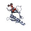
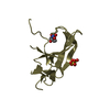
| #1: Protein | Mass: 12755.696 Da / Num. of mol.: 8 / Fragment: PDZ domain Source method: isolated from a genetically manipulated source Source: (gene. exp.)  #2: Protein/peptide | Mass: 847.869 Da / Num. of mol.: 8 / Source method: obtained synthetically / Details: synthetic peptide / References: UniProt: O55043*PLUS #3: Water | ChemComp-HOH / |  Mass: 18.015 Da / Num. of mol.: 129 / Source method: isolated from a natural source / Formula: H2O Mass: 18.015 Da / Num. of mol.: 129 / Source method: isolated from a natural source / Formula: H2O |
|---|
-Experimental details
-Experiment
| Experiment | Method: X-RAY DIFFRACTION / Number of used crystals: 1 |
|---|
- Sample preparation
| Crystal | Density Matthews: 2.13 Å3/Da / Density % sol: 42.19 % |
|---|---|
| Crystal grow | Temperature: 294 K / Method: vapor diffusion, hanging drop / pH: 6.5 Details: 100mM MES, 12% ethanol, 10% ethylene glycol, pH 6.5, VAPOR DIFFUSION, HANGING DROP, temperature 294K |
-Data collection
| Diffraction | Mean temperature: 100 K |
|---|---|
| Diffraction source | Source: SYNCHROTRON / Site: PAL/PLS  / Beamline: 4A / Wavelength: 1 Å / Beamline: 4A / Wavelength: 1 Å |
| Detector | Detector: CCD |
| Radiation | Protocol: SINGLE WAVELENGTH / Monochromatic (M) / Laue (L): M / Scattering type: x-ray |
| Radiation wavelength | Wavelength: 1 Å / Relative weight: 1 |
| Reflection | Resolution: 2.7→30 Å / Num. all: 24385 / Num. obs: 24385 / % possible obs: 94 % / Observed criterion σ(F): 0 / Observed criterion σ(I): 0 / Biso Wilson estimate: 71.03 Å2 |
| Reflection shell | Resolution: 2.7→2.8 Å / % possible all: 89.8 |
- Processing
| Software |
| ||||||||||||||||||||||||||||||||||||||||||||||||||||||||||||||||||||||||||||||||||||||||||||||||||||||||||||
|---|---|---|---|---|---|---|---|---|---|---|---|---|---|---|---|---|---|---|---|---|---|---|---|---|---|---|---|---|---|---|---|---|---|---|---|---|---|---|---|---|---|---|---|---|---|---|---|---|---|---|---|---|---|---|---|---|---|---|---|---|---|---|---|---|---|---|---|---|---|---|---|---|---|---|---|---|---|---|---|---|---|---|---|---|---|---|---|---|---|---|---|---|---|---|---|---|---|---|---|---|---|---|---|---|---|---|---|---|---|
| Refinement | Method to determine structure: MOLECULAR REPLACEMENT / Resolution: 2.71→21.3 Å / Cor.coef. Fo:Fc: 0.8992 / Cor.coef. Fo:Fc free: 0.8467 / Occupancy max: 1 / Occupancy min: 1 / Cross valid method: THROUGHOUT / σ(F): 0
| ||||||||||||||||||||||||||||||||||||||||||||||||||||||||||||||||||||||||||||||||||||||||||||||||||||||||||||
| Displacement parameters | Biso max: 158.44 Å2 / Biso mean: 69.24 Å2 / Biso min: 19.89 Å2
| ||||||||||||||||||||||||||||||||||||||||||||||||||||||||||||||||||||||||||||||||||||||||||||||||||||||||||||
| Refine analyze | Luzzati coordinate error obs: 0.379 Å | ||||||||||||||||||||||||||||||||||||||||||||||||||||||||||||||||||||||||||||||||||||||||||||||||||||||||||||
| Refinement step | Cycle: LAST / Resolution: 2.71→21.3 Å
| ||||||||||||||||||||||||||||||||||||||||||||||||||||||||||||||||||||||||||||||||||||||||||||||||||||||||||||
| Refine LS restraints |
| ||||||||||||||||||||||||||||||||||||||||||||||||||||||||||||||||||||||||||||||||||||||||||||||||||||||||||||
| LS refinement shell | Resolution: 2.71→2.83 Å / Total num. of bins used: 12
| ||||||||||||||||||||||||||||||||||||||||||||||||||||||||||||||||||||||||||||||||||||||||||||||||||||||||||||
| Refinement TLS params. | Method: refined / Origin x: 35.8438 Å / Origin y: 94.4871 Å / Origin z: 93.7995 Å
| ||||||||||||||||||||||||||||||||||||||||||||||||||||||||||||||||||||||||||||||||||||||||||||||||||||||||||||
| Refinement TLS group | Selection details: { A|656 - A|757 } |
