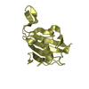
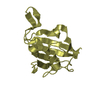
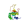
Mass: 10533.178 Da / Num. of mol.: 1 / Fragment: residues 309-400 Source method: isolated from a genetically manipulated source Source: (gene. exp.) Gallus gallus (chicken) / Gene: TLN1, TLN / Production host: Escherichia coli (E. coli) / Strain (production host): Bl21 / References: UniProt: P54939
#2: Protein/peptide
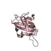
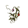
Layilin
Mass: 1818.938 Da / Num. of mol.: 1 / Fragment: residues 367-381 / Source method: obtained synthetically Details: The peptide is the C-terminal layilin fragment naturally found in Mus musculus. References: UniProt: Q8C351
-
Experimental details
-
Experiment
Experiment
Method: SOLUTION NMR
NMR experiment
Conditions-ID
Experiment-ID
Solution-ID
Type
1
1
1
3D 1H-13C filtered NOESY
1
2
1
3D 1H-15N filtered NOESY
1
3
1
2D 1H-1H filtered NOESY
NMR details
Text: The structure was determined using isotope-filtered experiments
-
Sample preparation
Details
Contents: 1 mM [U-13C; U-15N] protein, 3.5 mM peptide, 100 mM sodium chloride, 50 mM sodium phosphate, 90% H2O/10% D2O Solvent system: 90% H2O/10% D2O
Method: simulated annealing, molecular dynamics / Software ordinal: 1 Details: Structure of the complex calculated using 127 intermolecular NOE and 155 NOEs within the peptide. Final structures refined in water.
NMR representative
Selection criteria: lowest energy
NMR ensemble
Conformer selection criteria: structures with the lowest energy Conformers calculated total number: 200 / Conformers submitted total number: 20
+
About Yorodumi
-
News
-
Feb 9, 2022. New format data for meta-information of EMDB entries
New format data for meta-information of EMDB entries
Version 3 of the EMDB header file is now the official format.
The previous official version 1.9 will be removed from the archive.
In the structure databanks used in Yorodumi, some data are registered as the other names, "COVID-19 virus" and "2019-nCoV". Here are the details of the virus and the list of structure data.
Jan 31, 2019. EMDB accession codes are about to change! (news from PDBe EMDB page)
EMDB accession codes are about to change! (news from PDBe EMDB page)
The allocation of 4 digits for EMDB accession codes will soon come to an end. Whilst these codes will remain in use, new EMDB accession codes will include an additional digit and will expand incrementally as the available range of codes is exhausted. The current 4-digit format prefixed with “EMD-” (i.e. EMD-XXXX) will advance to a 5-digit format (i.e. EMD-XXXXX), and so on. It is currently estimated that the 4-digit codes will be depleted around Spring 2019, at which point the 5-digit format will come into force.
The EM Navigator/Yorodumi systems omit the EMD- prefix.
Related info.:Q: What is EMD? / ID/Accession-code notation in Yorodumi/EM Navigator
Yorodumi is a browser for structure data from EMDB, PDB, SASBDB, etc.
This page is also the successor to EM Navigator detail page, and also detail information page/front-end page for Omokage search.
The word "yorodu" (or yorozu) is an old Japanese word meaning "ten thousand". "mi" (miru) is to see.
Related info.:EMDB / PDB / SASBDB / Comparison of 3 databanks / Yorodumi Search / Aug 31, 2016. New EM Navigator & Yorodumi / Yorodumi Papers / Jmol/JSmol / Function and homology information / Changes in new EM Navigator and Yorodumi
 Movie
Movie Controller

 Yorodumi
 Open data
 Basic information
 Components
 Keywords
 Function and homology information
 Authors
 Citation
 Structure visualization
 Downloads & links
 Other downloads


 PDBj
PDBj


 Assembly
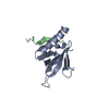

 Sample preparation
 Processing