 Movie
Movie Controller

[English] 日本語
 Yorodumi
- PDB-3q83: Crystal structure of Staphylococcus aureus nucleoside diphosphate... -
+ Open data

- Basic information
| Entry | Database: PDB / ID: 3q83 | ||||||
|---|---|---|---|---|---|---|---|
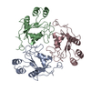
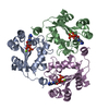
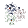
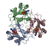
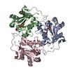
| Title | Crystal structure of Staphylococcus aureus nucleoside diphosphate kinase | ||||||
 Components Components | Nucleoside diphosphate kinase | ||||||
 Keywords Keywords | TRANSFERASE / Ferridoxin fold / alpha-beta protein family / Nucleoside diphosphate kinases (NDKs) catalyze the transfer of a gamma phosphate from nucleoside triphosphates to nucleoside diphosphate / Nucleotide binding / Metal binding / Phosphorylation / Kinase / Nucleotide metabolism | ||||||
| Function / homology |  Function and homology information Function and homology informationnucleoside-diphosphate kinase / UTP biosynthetic process / CTP biosynthetic process / nucleoside diphosphate kinase activity / GTP biosynthetic process / ATP binding / metal ion binding / cytoplasm Similarity search - Function | ||||||
| Biological species |   Staphylococcus aureus subsp. aureus (bacteria) Staphylococcus aureus subsp. aureus (bacteria) | ||||||
| Method |  X-RAY DIFFRACTION / SYNCHROTRON / MOLECULAR REPLACEMENT / Resolution: 2.5 Å X-RAY DIFFRACTION / SYNCHROTRON / MOLECULAR REPLACEMENT / Resolution: 2.5 Å | ||||||
 Authors Authors | Srivastava, S.K. / Rajasree, K. / Gopal, B. | ||||||
 Citation Citation | Journal: Biochim.Biophys.Acta / Year: 2011 Title: Conformational basis for substrate recognition and regulation of catalytic activity in Staphylococcus aureus nucleoside di-phosphate kinase. Authors: Srivastava, S.K. / Rajasree, K. / Gopal, B. | ||||||
| History |
|
- Structure visualization
| Structure viewer | Molecule: MolmilJmol/JSmol |
|---|
- Downloads & links
-Download
| PDBx/mmCIF format | 3q83.cif.gz | 186 KB | Display | PDBx/mmCIF format |
|---|---|---|---|---|
| PDB format | pdb3q83.ent.gz | 150.2 KB | Display | PDB format |
| PDBx/mmJSON format | 3q83.json.gz | Tree view | PDBx/mmJSON format | |
| Others |  Other downloads Other downloads |
-Validation report
| Arichive directory | https://data.pdbj.org/pub/pdb/validation_reports/q8/3q83ftp://data.pdbj.org/pub/pdb/validation_reports/q8/3q83 | HTTPS FTP |
|---|
-Related structure data
| Related structure data |  3q86C  3q89C  3q8uC  3q8vC  3q8yC  1jxvS C: citing same article ( S: Starting model for refinement |
|---|---|
| Similar structure data |


-Links
 PDBj
PDBj- Assembly
| Deposited unit | 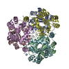
| ||||||||||||||||||||||||||||||||||||||||||
|---|---|---|---|---|---|---|---|---|---|---|---|---|---|---|---|---|---|---|---|---|---|---|---|---|---|---|---|---|---|---|---|---|---|---|---|---|---|---|---|---|---|---|---|
| 1 | 
| ||||||||||||||||||||||||||||||||||||||||||
| 2 | 
| ||||||||||||||||||||||||||||||||||||||||||
| 3 |
| ||||||||||||||||||||||||||||||||||||||||||
| Unit cell |
| ||||||||||||||||||||||||||||||||||||||||||
| Noncrystallographic symmetry (NCS) | NCS domain:
NCS domain segments: Component-ID: 1 / Ens-ID: 1 / Beg auth comp-ID: MET / Beg label comp-ID: MET / End auth comp-ID: GLU / End label comp-ID: GLU / Refine code: 2 / Auth seq-ID: 1 - 149 / Label seq-ID: 1 - 149
|
-Components
| #1: Protein | Mass: 17663.873 Da / Num. of mol.: 6 Source method: isolated from a genetically manipulated source Details: genomic DNA Source: (gene. exp.) Staphylococcus aureus subsp. aureus (bacteria)Strain: COL / Gene: ndk, SACOL1509 / Plasmid: pET22b / Production host: #2: Water | ChemComp-HOH / |  Mass: 18.015 Da / Num. of mol.: 429 / Source method: isolated from a natural source / Formula: H2O Mass: 18.015 Da / Num. of mol.: 429 / Source method: isolated from a natural source / Formula: H2O |
|---|
-Experimental details
-Experiment
| Experiment | Method: X-RAY DIFFRACTION / Number of used crystals: 1 |
|---|
- Sample preparation
| Crystal | Density Matthews: 2.02 Å3/Da / Density % sol: 39.16 % |
|---|---|
| Crystal grow | Temperature: 298 K / Method: micobatch crystallization / pH: 4.6 Details: 0.1M MgCl2, 0.1M Na-Acetate, pH 4.6, 20% PEG 2000, Micobatch Crystallization, temperature 298K |
-Data collection
| Diffraction | Mean temperature: 100 K |
|---|---|
| Diffraction source | Source: SYNCHROTRON / Site: ESRF  / Beamline: BM14 / Wavelength: 0.9537 Å / Beamline: BM14 / Wavelength: 0.9537 Å |
| Detector | Type: MARMOSAIC 225 mm CCD / Detector: CCD / Date: Nov 1, 2009 / Details: Grazing angle 2.8 mrad |
| Radiation | Monochromator: Si(111) monochromator / Protocol: SINGLE WAVELENGTH / Monochromatic (M) / Laue (L): M / Scattering type: x-ray |
| Radiation wavelength | Wavelength: 0.9537 Å / Relative weight: 1 |
| Reflection | Resolution: 2.5→72.36 Å / Num. obs: 30434 / % possible obs: 99.8 % / Observed criterion σ(F): 1 / Observed criterion σ(I): 0 / Redundancy: 5.5 % / Biso Wilson estimate: 30.8 Å2 / Rmerge(I) obs: 0.156 / Net I/σ(I): 7.5 |
| Reflection shell | Resolution: 2.5→2.64 Å / Redundancy: 5.5 % / Rmerge(I) obs: 0.38 / Mean I/σ(I) obs: 3.8 / Num. unique all: 4378 / % possible all: 100 |
- Processing
| Software |
| |||||||||||||||||||||||||||||||||||||||||||||||||||||||||||||||||||||||||||||||||||||||||||||||||||||||||||||||||||||||||||||
|---|---|---|---|---|---|---|---|---|---|---|---|---|---|---|---|---|---|---|---|---|---|---|---|---|---|---|---|---|---|---|---|---|---|---|---|---|---|---|---|---|---|---|---|---|---|---|---|---|---|---|---|---|---|---|---|---|---|---|---|---|---|---|---|---|---|---|---|---|---|---|---|---|---|---|---|---|---|---|---|---|---|---|---|---|---|---|---|---|---|---|---|---|---|---|---|---|---|---|---|---|---|---|---|---|---|---|---|---|---|---|---|---|---|---|---|---|---|---|---|---|---|---|---|---|---|---|
| Refinement | Method to determine structure: MOLECULAR REPLACEMENT Starting model: PDB entry 1JXV Resolution: 2.5→45.81 Å / Cor.coef. Fo:Fc: 0.9 / Cor.coef. Fo:Fc free: 0.846 / SU B: 10.569 / SU ML: 0.238 / Cross valid method: THROUGHOUT / ESU R Free: 0.345 / Stereochemistry target values: MAXIMUM LIKELIHOOD / Details: HYDROGENS HAVE BEEN ADDED IN THE RIDING POSITIONS
| |||||||||||||||||||||||||||||||||||||||||||||||||||||||||||||||||||||||||||||||||||||||||||||||||||||||||||||||||||||||||||||
| Solvent computation | Ion probe radii: 0.8 Å / Shrinkage radii: 0.8 Å / VDW probe radii: 1.4 Å / Solvent model: MASK | |||||||||||||||||||||||||||||||||||||||||||||||||||||||||||||||||||||||||||||||||||||||||||||||||||||||||||||||||||||||||||||
| Displacement parameters | Biso mean: 16.18 Å2
| |||||||||||||||||||||||||||||||||||||||||||||||||||||||||||||||||||||||||||||||||||||||||||||||||||||||||||||||||||||||||||||
| Refine analyze | Luzzati coordinate error obs: 0.345 Å | |||||||||||||||||||||||||||||||||||||||||||||||||||||||||||||||||||||||||||||||||||||||||||||||||||||||||||||||||||||||||||||
| Refinement step | Cycle: LAST / Resolution: 2.5→45.81 Å
| |||||||||||||||||||||||||||||||||||||||||||||||||||||||||||||||||||||||||||||||||||||||||||||||||||||||||||||||||||||||||||||
| Refine LS restraints |
| |||||||||||||||||||||||||||||||||||||||||||||||||||||||||||||||||||||||||||||||||||||||||||||||||||||||||||||||||||||||||||||
| Refine LS restraints NCS | Dom-ID: 1 / Ens-ID: 1 / Refine-ID: X-RAY DIFFRACTION
| |||||||||||||||||||||||||||||||||||||||||||||||||||||||||||||||||||||||||||||||||||||||||||||||||||||||||||||||||||||||||||||
| LS refinement shell | Resolution: 2.5→2.565 Å / Total num. of bins used: 20
|
