 Movie
Movie Controller


+ Open data

- Basic information
| Entry | Database: PDB / ID: 2hvd | ||||||
|---|---|---|---|---|---|---|---|
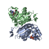
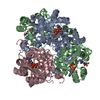
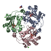
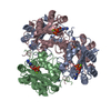
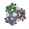
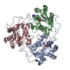
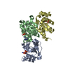
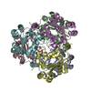
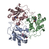
| Title | Human nucleoside diphosphate kinase A complexed with ADP | ||||||
 Components Components | Nucleoside diphosphate kinase A | ||||||
 Keywords Keywords | SIGNALING PROTEIN / TRANSFERASE / COMPLEX ADP | ||||||
| Function / homology |  Function and homology information Function and homology informationfarnesyl-diphosphate kinase / farnesyl diphosphate kinase activity / succinyl-CoA binding / ITP biosynthetic process / isoprenoid metabolic process / Hydrolases; Acting on ester bonds; Site specific endodeoxyribonucleases: cleavage is not sequence specific (deleted sub-subclass) / phosphotransferase activity, phosphate group as acceptor / DNA nuclease activity / acetyl-CoA catabolic process / nucleoside triphosphate biosynthetic process ...farnesyl-diphosphate kinase / farnesyl diphosphate kinase activity / succinyl-CoA binding / ITP biosynthetic process / isoprenoid metabolic process / Hydrolases; Acting on ester bonds; Site specific endodeoxyribonucleases: cleavage is not sequence specific (deleted sub-subclass) / phosphotransferase activity, phosphate group as acceptor / DNA nuclease activity / acetyl-CoA catabolic process / nucleoside triphosphate biosynthetic process / acetyl-CoA binding / nucleoside diphosphate metabolic process / Ribavirin ADME / coenzyme A binding / protein histidine kinase activity / regulation of fatty acid biosynthetic process / apoptotic DNA fragmentation / nucleoside-diphosphate kinase / 3'-5'-DNA exonuclease activity / Interconversion of nucleotide di- and triphosphates / UTP biosynthetic process / CTP biosynthetic process / protein hexamerization / Azathioprine ADME / DNA catabolic process / nucleoside diphosphate kinase activity / GTP biosynthetic process / Hydrolases; Acting on ester bonds; Exodeoxyribonucleases producing 5'-phosphomonoesters / histidine kinase / ribosomal small subunit binding / lactation / positive regulation of epithelial cell proliferation / DNA endonuclease activity / ADP binding / ruffle membrane / endocytosis / kinase activity / GDP binding / nervous system development / regulation of apoptotic process / early endosome / cell differentiation / non-specific serine/threonine protein kinase / negative regulation of cell population proliferation / magnesium ion binding / DNA binding / RNA binding / extracellular exosome / ATP binding / membrane / identical protein binding / nucleus / cytoplasm / cytosol Similarity search - Function | ||||||
| Biological species |  Homo sapiens (human) Homo sapiens (human) | ||||||
| Method |  X-RAY DIFFRACTION / SYNCHROTRON / MOLECULAR REPLACEMENT / Resolution: 2.15 Å X-RAY DIFFRACTION / SYNCHROTRON / MOLECULAR REPLACEMENT / Resolution: 2.15 Å | ||||||
 Authors Authors | Giraud, M.-F. / Georgescauld, F. / Lascu, I. / Dautant, A. | ||||||
 Citation Citation | Journal: J.Bioenerg.Biomembr. / Year: 2006 Title: Crystal Structures of S120G Mutant and Wild Type of Human Nucleoside Diphosphate Kinase A in Complex with ADP Authors: Giraud, M.-F. / Georgescauld, F. / Lascu, I. / Dautant, A. #1: Journal: Proteins / Year: 2002Title: Crystal structure of human nucleoside diphosphate kinase A, a metastasis suppressor Authors: Min, K. / Song, H.K. / Chang, C. / Kim, S.Y. / Lee, K.J. / Suh, S.W. #2: Journal: J.Mol.Biol. / Year: 2003Title: Nucleotide binding to nucleoside diphosphate kinases: X-ray structure of human NDPK-A in complex with ADP and comparison to protein kinases Authors: Chen, Y. / Gallois-Montbrun, S. / Schneider, B. / Deville-Bonne, D. / Janin, J. | ||||||
| History |
|
- Structure visualization
| Structure viewer | Molecule: MolmilJmol/JSmol |
|---|
- Downloads & links
-Download
| PDBx/mmCIF format | 2hvd.cif.gz | 106.9 KB | Display | PDBx/mmCIF format |
|---|---|---|---|---|
| PDB format | pdb2hvd.ent.gz | 83.3 KB | Display | PDB format |
| PDBx/mmJSON format | 2hvd.json.gz | Tree view | PDBx/mmJSON format | |
| Others |  Other downloads Other downloads |
-Validation report
| Arichive directory | https://data.pdbj.org/pub/pdb/validation_reports/hv/2hvdftp://data.pdbj.org/pub/pdb/validation_reports/hv/2hvd | HTTPS FTP |
|---|
-Related structure data
| Related structure data | 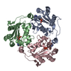 2hveC  1ucnS C: citing same article ( S: Starting model for refinement |
|---|---|
| Similar structure data |


-Links
 PDBj
PDBj


- Assembly
| Deposited unit | 
| |||||||||||||||
|---|---|---|---|---|---|---|---|---|---|---|---|---|---|---|---|---|
| 1 | 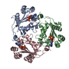
| |||||||||||||||
| Unit cell |
| |||||||||||||||
| Components on special symmetry positions |
|
-Components
| #1: Protein | Mass: 17170.721 Da / Num. of mol.: 3 Source method: isolated from a genetically manipulated source Source: (gene. exp.) Homo sapiens (human) / Gene: NME1, NDPKA, NM23 / Plasmid: PET-21 / Production host:  #2: Chemical |   Mass: 427.201 Da / Num. of mol.: 3 / Source method: obtained synthetically / Formula: C10H15N5O10P2 / Comment: ADP, energy-carrying molecule*YM Mass: 427.201 Da / Num. of mol.: 3 / Source method: obtained synthetically / Formula: C10H15N5O10P2 / Comment: ADP, energy-carrying molecule*YM#3: Water | ChemComp-HOH / |  Mass: 18.015 Da / Num. of mol.: 190 / Source method: isolated from a natural source / Formula: H2O Mass: 18.015 Da / Num. of mol.: 190 / Source method: isolated from a natural source / Formula: H2OHas protein modification | Y | |
|---|
-Experimental details
-Experiment
| Experiment | Method: X-RAY DIFFRACTION / Number of used crystals: 1 |
|---|
- Sample preparation
| Crystal | Density Matthews: 2.85 Å3/Da / Density % sol: 56.5 % |
|---|---|
| Crystal grow | Temperature: 293 K / Method: vapor diffusion, sitting drop / pH: 6 Details: 2.4 M AMMONIUM SULPHATE,10 MM ADP, 20 MM MGCL2, 4 MM DTT, 0.1 M MES, pH 6.0, VAPOR DIFFUSION, SITTING DROP, temperature 293K |
-Data collection
| Diffraction | Mean temperature: 100 K |
|---|---|
| Diffraction source | Source: SYNCHROTRON / Site: ESRF  / Beamline: BM30A / Wavelength: 0.973 / Wavelength: 0.973 Å / Beamline: BM30A / Wavelength: 0.973 / Wavelength: 0.973 Å |
| Detector | Type: MARRESEARCH / Detector: CCD / Date: Mar 7, 2005 |
| Radiation | Protocol: SINGLE WAVELENGTH / Monochromatic (M) / Laue (L): M / Scattering type: x-ray |
| Radiation wavelength | Wavelength: 0.973 Å / Relative weight: 1 |
| Reflection | Resolution: 2.15→17.65 Å / Num. all: 30827 / Num. obs: 30827 / % possible obs: 97.2 % / Observed criterion σ(F): 0 / Observed criterion σ(I): 0 / Redundancy: 3.2 % / Biso Wilson estimate: 39.084 Å2 / Rsym value: 0.08 / Net I/σ(I): 10.2 |
| Reflection shell | Resolution: 2.15→2.27 Å / Redundancy: 3.1 % / Mean I/σ(I) obs: 3.1 / Rsym value: 0.52 / % possible all: 99 |
- Processing
| Software |
| ||||||||||||||||||||||||||||||||||||||||||||||||||||||||||||||||||||||||||||||||||||||||||||||||||||||||||||||||||||||||||||||||||
|---|---|---|---|---|---|---|---|---|---|---|---|---|---|---|---|---|---|---|---|---|---|---|---|---|---|---|---|---|---|---|---|---|---|---|---|---|---|---|---|---|---|---|---|---|---|---|---|---|---|---|---|---|---|---|---|---|---|---|---|---|---|---|---|---|---|---|---|---|---|---|---|---|---|---|---|---|---|---|---|---|---|---|---|---|---|---|---|---|---|---|---|---|---|---|---|---|---|---|---|---|---|---|---|---|---|---|---|---|---|---|---|---|---|---|---|---|---|---|---|---|---|---|---|---|---|---|---|---|---|---|---|
| Refinement | Method to determine structure: MOLECULAR REPLACEMENT Starting model: PDB ENTRY 1UCN Resolution: 2.15→17.65 Å / Cor.coef. Fo:Fc: 0.926 / Cor.coef. Fo:Fc free: 0.894 / SU B: 7.653 / SU ML: 0.197 / Cross valid method: THROUGHOUT / σ(F): 0 / ESU R: 0.272 / ESU R Free: 0.229 / Stereochemistry target values: MAXIMUM LIKELIHOOD / Details: HYDROGENS HAVE BEEN ADDED IN THE RIDING POSITIONS
| ||||||||||||||||||||||||||||||||||||||||||||||||||||||||||||||||||||||||||||||||||||||||||||||||||||||||||||||||||||||||||||||||||
| Solvent computation | Ion probe radii: 0.8 Å / Shrinkage radii: 0.8 Å / VDW probe radii: 1.2 Å / Solvent model: BABINET MODEL WITH MASK | ||||||||||||||||||||||||||||||||||||||||||||||||||||||||||||||||||||||||||||||||||||||||||||||||||||||||||||||||||||||||||||||||||
| Displacement parameters | Biso mean: 42.196 Å2
| ||||||||||||||||||||||||||||||||||||||||||||||||||||||||||||||||||||||||||||||||||||||||||||||||||||||||||||||||||||||||||||||||||
| Refinement step | Cycle: LAST / Resolution: 2.15→17.65 Å
| ||||||||||||||||||||||||||||||||||||||||||||||||||||||||||||||||||||||||||||||||||||||||||||||||||||||||||||||||||||||||||||||||||
| Refine LS restraints |
| ||||||||||||||||||||||||||||||||||||||||||||||||||||||||||||||||||||||||||||||||||||||||||||||||||||||||||||||||||||||||||||||||||
| LS refinement shell | Resolution: 2.15→2.206 Å / Total num. of bins used: 20
|
