- PDB-1ucn: X-ray structure of human nucleoside diphosphate kinase A complexe... -
+
Open data
ID or keywords:
Loading...
-
Basic information
Entry
Database: PDB / ID: 1ucn
Title
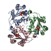
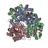
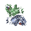
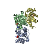
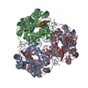
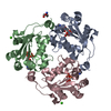
X-ray structure of human nucleoside diphosphate kinase A complexed with ADP at 2 A resolution
Components
nucleoside diphosphate kinase A
Keywords
TRANSFERASE / mutant complexe ADP
Function / homology
Function and homology information
farnesyl-diphosphate kinase / farnesyl diphosphate kinase activity / succinyl-CoA binding / ITP biosynthetic process / isoprenoid metabolic process / Hydrolases; Acting on ester bonds; Site specific endodeoxyribonucleases: cleavage is not sequence specific (deleted sub-subclass) / phosphotransferase activity, phosphate group as acceptor / DNA nuclease activity / acetyl-CoA catabolic process / nucleoside triphosphate biosynthetic process ...farnesyl-diphosphate kinase / farnesyl diphosphate kinase activity / succinyl-CoA binding / ITP biosynthetic process / isoprenoid metabolic process / Hydrolases; Acting on ester bonds; Site specific endodeoxyribonucleases: cleavage is not sequence specific (deleted sub-subclass) / phosphotransferase activity, phosphate group as acceptor / DNA nuclease activity / acetyl-CoA catabolic process / nucleoside triphosphate biosynthetic process / acetyl-CoA binding / nucleoside diphosphate metabolic process / Ribavirin ADME / protein histidine kinase activity / coenzyme A binding / regulation of fatty acid biosynthetic process / apoptotic DNA fragmentation / nucleoside-diphosphate kinase / 3'-5'-DNA exonuclease activity / Interconversion of nucleotide di- and triphosphates / UTP biosynthetic process / CTP biosynthetic process / protein hexamerization / Azathioprine ADME / DNA catabolic process / nucleoside diphosphate kinase activity / GTP biosynthetic process / Hydrolases; Acting on ester bonds; Exodeoxyribonucleases producing 5'-phosphomonoesters / histidine kinase / ribosomal small subunit binding / positive regulation of epithelial cell proliferation / DNA endonuclease activity / ADP binding / ruffle membrane / endocytosis / kinase activity / GDP binding / nervous system development / regulation of apoptotic process / early endosome / cell differentiation / non-specific serine/threonine protein kinase / negative regulation of cell population proliferation / protein serine kinase activity / protein serine/threonine kinase activity / magnesium ion binding / DNA binding / RNA binding / extracellular exosome / ATP binding / membrane / identical protein binding / nucleus / cytosol / cytoplasm Similarity search - Function
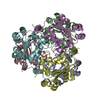
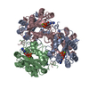
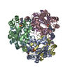
The second part of the biological assembly is generated by the two fold axis: x,y,z * -x,-y,1/2+z * 1/2-y,1/2+x,1/4+z * 1/2+y,1/2-x,3/4+z * 1/2-x,1/2+y,1/4-z * 1/2+x,1/2-y,3/4-z * y,x,-z * -y,-x,1/2-z *
-
Components
-
Protein , 1 types, 3 molecules ABC
#1: Protein
nucleosidediphosphatekinaseA
Mass: 17128.660 Da / Num. of mol.: 3 / Mutation: H118G, F60W Source method: isolated from a genetically manipulated source Source: (gene. exp.) Homo sapiens (human) / Plasmid: pJC20 / Production host: Escherichia coli (E. coli) / References: UniProt: P15531, nucleoside-diphosphate kinase
In the structure databanks used in Yorodumi, some data are registered as the other names, "COVID-19 virus" and "2019-nCoV". Here are the details of the virus and the list of structure data.
Jan 31, 2019. EMDB accession codes are about to change! (news from PDBe EMDB page)
EMDB accession codes are about to change! (news from PDBe EMDB page)
The allocation of 4 digits for EMDB accession codes will soon come to an end. Whilst these codes will remain in use, new EMDB accession codes will include an additional digit and will expand incrementally as the available range of codes is exhausted. The current 4-digit format prefixed with “EMD-” (i.e. EMD-XXXX) will advance to a 5-digit format (i.e. EMD-XXXXX), and so on. It is currently estimated that the 4-digit codes will be depleted around Spring 2019, at which point the 5-digit format will come into force.
The EM Navigator/Yorodumi systems omit the EMD- prefix.
Related info.:Q: What is EMD? / ID/Accession-code notation in Yorodumi/EM Navigator
Yorodumi is a browser for structure data from EMDB, PDB, SASBDB, etc.
This page is also the successor to EM Navigator detail page, and also detail information page/front-end page for Omokage search.
The word "yorodu" (or yorozu) is an old Japanese word meaning "ten thousand". "mi" (miru) is to see.
Related info.:EMDB / PDB / SASBDB / Comparison of 3 databanks / Yorodumi Search / Aug 31, 2016. New EM Navigator & Yorodumi / Yorodumi Papers / Jmol/JSmol / Function and homology information / Changes in new EM Navigator and Yorodumi
 Movie
Movie Controller

 Yorodumi
 Open data
 Basic information
 Components
 Keywords
 Function and homology information
 Homo sapiens (human)
 X-RAY DIFFRACTION /
  Authors
 Citation
 Structure visualization
 Downloads & links
 Other downloads
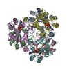


 PDBj
PDBj


 Assembly


 Mass: 94.971 Da / Num. of mol.: 3 / Source method: obtained synthetically / Formula: PO4
 Mass: 40.078 Da / Num. of mol.: 5 / Source method: obtained synthetically / Formula: Ca
 Mass: 427.201 Da / Num. of mol.: 3 / Source method: obtained synthetically / Formula: C10H15N5O10P2 / Comment: ADP, energy-carrying molecule*YM
 Mass: 122.143 Da / Num. of mol.: 1 / Source method: obtained synthetically / Formula: C4H12NO3 / Comment: pH buffer*YM
 Sample preparation
 / Beamline: ID14-1 / Wavelength: 0.934 / Wavelength: 0.934 Å
 Processing