 Movie
Movie Controller

[English] 日本語
 Yorodumi
- PDB-2dxd: Crystal structure of nucleoside diphosphate kinase in complex wit... -
+ Open data

- Basic information
| Entry | Database: PDB / ID: 2dxd | |||||||||
|---|---|---|---|---|---|---|---|---|---|---|
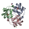
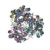
| Title | Crystal structure of nucleoside diphosphate kinase in complex with ATP analog | |||||||||
 Components Components | Nucleoside diphosphate kinase | |||||||||
 Keywords Keywords | TRANSFERASE / nucleoside binding / kinase / Structural Genomics / NPPSFA / National Project on Protein Structural and Functional Analyses / RIKEN Structural Genomics/Proteomics Initiative / RSGI | |||||||||
| Function / homology |  Function and homology information Function and homology informationnucleoside-diphosphate kinase / UTP biosynthetic process / CTP biosynthetic process / nucleoside diphosphate kinase activity / GTP biosynthetic process / ATP binding / metal ion binding / cytoplasm Similarity search - Function | |||||||||
| Biological species |   Pyrococcus horikoshii (archaea) Pyrococcus horikoshii (archaea) | |||||||||
| Method |  X-RAY DIFFRACTION / MOLECULAR REPLACEMENT / Resolution: 1.77 Å X-RAY DIFFRACTION / MOLECULAR REPLACEMENT / Resolution: 1.77 Å | |||||||||
 Authors Authors | Kato-Murayama, M. / Murayama, K. / Terada, T. / Shirouzu, M. / Yokoyama, S. / RIKEN Structural Genomics/Proteomics Initiative (RSGI) | |||||||||
 Citation Citation | Journal: To be Published Title: Crystal structure of nucleoside diphosphate kinase in complex with ATP analog Authors: Kato-Murayama, M. / Murayama, K. / Terada, T. / Shirouzu, M. / Yokoyama, S. | |||||||||
| History |
|
- Structure visualization
| Structure viewer | Molecule: MolmilJmol/JSmol |
|---|
- Downloads & links
-Download
| PDBx/mmCIF format | 2dxd.cif.gz | 86.6 KB | Display | PDBx/mmCIF format |
|---|---|---|---|---|
| PDB format | pdb2dxd.ent.gz | 64 KB | Display | PDB format |
| PDBx/mmJSON format | 2dxd.json.gz | Tree view | PDBx/mmJSON format | |
| Others |  Other downloads Other downloads |
-Validation report
| Arichive directory | https://data.pdbj.org/pub/pdb/validation_reports/dx/2dxdftp://data.pdbj.org/pub/pdb/validation_reports/dx/2dxd | HTTPS FTP |
|---|
-Related structure data
| Related structure data |  2cwkS S: Starting model for refinement |
|---|---|
| Similar structure data | |
| Other databases |


-Links
 PDBj
PDBj


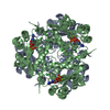
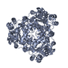
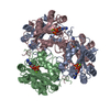
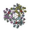
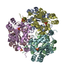
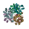
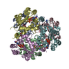
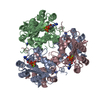
- Assembly
| Deposited unit | 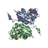
| ||||||||
|---|---|---|---|---|---|---|---|---|---|
| 1 | 
| ||||||||
| 2 | x 6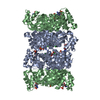
| ||||||||
| Unit cell |
|
-Components
| #1: Protein | Mass: 18342.336 Da / Num. of mol.: 2 Source method: isolated from a genetically manipulated source Source: (gene. exp.) Pyrococcus horikoshii (strain ATCC 700860 / DSM 12428 / JCM 9974 / NBRC 100139 / OT-3) (archaea)Species: Pyrococcus horikoshii Strain: ATCC 700860 / DSM 12428 / JCM 9974 / NBRC 100139 / OT-3 Gene: ndk, PH0698 / Plasmid: pET11a / Production host:  #2: Chemical |   Mass: 35.453 Da / Num. of mol.: 2 / Source method: obtained synthetically / Formula: Cl Mass: 35.453 Da / Num. of mol.: 2 / Source method: obtained synthetically / Formula: Cl#3: Chemical |   Mass: 347.221 Da / Num. of mol.: 2 / Source method: obtained synthetically / Formula: C10H14N5O7P / Comment: AMP*YM Mass: 347.221 Da / Num. of mol.: 2 / Source method: obtained synthetically / Formula: C10H14N5O7P / Comment: AMP*YM#4: Water | ChemComp-HOH / |  Mass: 18.015 Da / Num. of mol.: 434 / Source method: isolated from a natural source / Formula: H2O Mass: 18.015 Da / Num. of mol.: 434 / Source method: isolated from a natural source / Formula: H2O |
|---|
-Experimental details
-Experiment
| Experiment | Method: X-RAY DIFFRACTION / Number of used crystals: 1 |
|---|
- Sample preparation
| Crystal | Density Matthews: 3.4 Å3/Da / Density % sol: 63.86 % |
|---|---|
| Crystal grow | Temperature: 293 K / Method: vapor diffusion, hanging drop / pH: 7.5 Details: Na Citrate, Bicine, 10mM ANP, pH 7.5, VAPOR DIFFUSION, HANGING DROP, temperature 293K |
-Data collection
| Diffraction | Mean temperature: 100 K |
|---|---|
| Diffraction source | Source: ROTATING ANODE / Type: RIGAKU / Wavelength: 1.5418 Å |
| Detector | Type: RIGAKU RAXIS IV / Detector: IMAGE PLATE / Date: Dec 27, 2005 |
| Radiation | Monochromator: conforcal mirror / Protocol: SINGLE WAVELENGTH / Monochromatic (M) / Laue (L): M / Scattering type: x-ray |
| Radiation wavelength | Wavelength: 1.5418 Å / Relative weight: 1 |
| Reflection | Resolution: 1.77→87.71 Å / Num. obs: 48556 / % possible obs: 99.4 % / Observed criterion σ(I): -3 / Redundancy: 10.8 % / Biso Wilson estimate: 19.1 Å2 / Rsym value: 0.062 / Net I/σ(I): 34.5 |
| Reflection shell | Resolution: 1.77→1.83 Å / Rsym value: 0.317 / % possible all: 94.3 |
- Processing
| Software |
| ||||||||||||||||||||||||||||||||||||||||||||||||||||||||||||||||||||||||||||||||
|---|---|---|---|---|---|---|---|---|---|---|---|---|---|---|---|---|---|---|---|---|---|---|---|---|---|---|---|---|---|---|---|---|---|---|---|---|---|---|---|---|---|---|---|---|---|---|---|---|---|---|---|---|---|---|---|---|---|---|---|---|---|---|---|---|---|---|---|---|---|---|---|---|---|---|---|---|---|---|---|---|---|
| Refinement | Method to determine structure: MOLECULAR REPLACEMENT Starting model: PDB ENTRY 2CWK Resolution: 1.77→37.15 Å / Rfactor Rfree error: 0.003 / Data cutoff high absF: 3225959.86 / Data cutoff low absF: 0 / Isotropic thermal model: RESTRAINED / Cross valid method: THROUGHOUT / σ(F): 0
| ||||||||||||||||||||||||||||||||||||||||||||||||||||||||||||||||||||||||||||||||
| Solvent computation | Solvent model: FLAT MODEL / Bsol: 40.8777 Å2 / ksol: 0.387272 e/Å3 | ||||||||||||||||||||||||||||||||||||||||||||||||||||||||||||||||||||||||||||||||
| Displacement parameters | Biso mean: 20.2 Å2
| ||||||||||||||||||||||||||||||||||||||||||||||||||||||||||||||||||||||||||||||||
| Refine analyze |
| ||||||||||||||||||||||||||||||||||||||||||||||||||||||||||||||||||||||||||||||||
| Refinement step | Cycle: LAST / Resolution: 1.77→37.15 Å
| ||||||||||||||||||||||||||||||||||||||||||||||||||||||||||||||||||||||||||||||||
| Refine LS restraints |
| ||||||||||||||||||||||||||||||||||||||||||||||||||||||||||||||||||||||||||||||||
| LS refinement shell | Resolution: 1.77→1.85 Å / Rfactor Rfree error: 0.01 / Total num. of bins used: 8
| ||||||||||||||||||||||||||||||||||||||||||||||||||||||||||||||||||||||||||||||||
| Xplor file |
|
