 Movie
Movie Controller


+ Open data

- Basic information
| Entry | Database: PDB / ID: 1jxv | ||||||
|---|---|---|---|---|---|---|---|
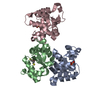
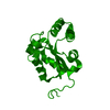
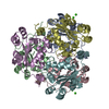
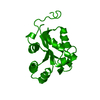
| Title | Crystal Structure of Human Nucleoside Diphosphate Kinase A | ||||||
 Components Components | Nucleoside Diphosphate Kinase A | ||||||
 Keywords Keywords | TRANSFERASE / kinase | ||||||
| Function / homology |  Function and homology information Function and homology informationfarnesyl-diphosphate kinase / farnesyl diphosphate kinase activity / succinyl-CoA binding / ITP biosynthetic process / isoprenoid metabolic process / Hydrolases; Acting on ester bonds; Site specific endodeoxyribonucleases: cleavage is not sequence specific (deleted sub-subclass) / phosphotransferase activity, phosphate group as acceptor / DNA nuclease activity / acetyl-CoA catabolic process / nucleoside triphosphate biosynthetic process ...farnesyl-diphosphate kinase / farnesyl diphosphate kinase activity / succinyl-CoA binding / ITP biosynthetic process / isoprenoid metabolic process / Hydrolases; Acting on ester bonds; Site specific endodeoxyribonucleases: cleavage is not sequence specific (deleted sub-subclass) / phosphotransferase activity, phosphate group as acceptor / DNA nuclease activity / acetyl-CoA catabolic process / nucleoside triphosphate biosynthetic process / acetyl-CoA binding / nucleoside diphosphate metabolic process / Ribavirin ADME / coenzyme A binding / protein histidine kinase activity / regulation of fatty acid biosynthetic process / apoptotic DNA fragmentation / nucleoside-diphosphate kinase / 3'-5'-DNA exonuclease activity / Interconversion of nucleotide di- and triphosphates / UTP biosynthetic process / CTP biosynthetic process / protein hexamerization / Azathioprine ADME / DNA catabolic process / nucleoside diphosphate kinase activity / GTP biosynthetic process / Hydrolases; Acting on ester bonds; Exodeoxyribonucleases producing 5'-phosphomonoesters / histidine kinase / ribosomal small subunit binding / lactation / positive regulation of epithelial cell proliferation / DNA endonuclease activity / ADP binding / ruffle membrane / endocytosis / kinase activity / GDP binding / nervous system development / regulation of apoptotic process / early endosome / cell differentiation / non-specific serine/threonine protein kinase / negative regulation of cell population proliferation / magnesium ion binding / DNA binding / RNA binding / extracellular exosome / ATP binding / membrane / identical protein binding / nucleus / cytosol / cytoplasm Similarity search - Function | ||||||
| Biological species |  Homo sapiens (human) Homo sapiens (human) | ||||||
| Method |  X-RAY DIFFRACTION / SYNCHROTRON / MOLECULAR REPLACEMENT / Resolution: 2.2 Å X-RAY DIFFRACTION / SYNCHROTRON / MOLECULAR REPLACEMENT / Resolution: 2.2 Å | ||||||
 Authors Authors | Min, K. / Song, H.K. / Chang, C. / Kim, S.Y. / Lee, K.J. / Suh, S.W. | ||||||
 Citation Citation | Journal: Proteins / Year: 2002 Title: Crystal structure of human nucleoside diphosphate kinase A, a metastasis suppressor. Authors: Min, K. / Song, H.K. / Chang, C. / Kim, S.Y. / Lee, K.J. / Suh, S.W. #1: Journal: Acta Crystallogr.,Sect.D / Year: 2000Title: Crystallization and preliminary X-ray crystallographic analysis of human nucleoside diphosphate kinase A Authors: Min, K. / Kim, S.Y. / Song, H.K. / Chang, C. / Cho, S.J. / Moon, J. / Yang, J.K. / Lee, J.Y. / Lee, K.J. / Suh, S.W. | ||||||
| History |
|
- Structure visualization
| Structure viewer | Molecule: MolmilJmol/JSmol |
|---|
- Downloads & links
-Download
| PDBx/mmCIF format | 1jxv.cif.gz | 184.2 KB | Display | PDBx/mmCIF format |
|---|---|---|---|---|
| PDB format | pdb1jxv.ent.gz | 149.2 KB | Display | PDB format |
| PDBx/mmJSON format | 1jxv.json.gz | Tree view | PDBx/mmJSON format | |
| Others |  Other downloads Other downloads |
-Validation report
| Arichive directory | https://data.pdbj.org/pub/pdb/validation_reports/jx/1jxvftp://data.pdbj.org/pub/pdb/validation_reports/jx/1jxv | HTTPS FTP |
|---|
-Related structure data
| Related structure data | 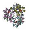 1nueS S: Starting model for refinement |
|---|---|
| Similar structure data |


-Links
 PDBj
PDBj


- Assembly
| Deposited unit | 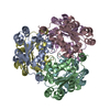
| ||||||||
|---|---|---|---|---|---|---|---|---|---|
| 1 |
| ||||||||
| Unit cell |
|
-Components
| #1: Protein | Mass: 17170.721 Da / Num. of mol.: 6 Source method: isolated from a genetically manipulated source Source: (gene. exp.) Homo sapiens (human) / Gene: nm23-H1 / Plasmid: pET-3c / Species (production host): Escherichia coli / Production host:  #2: Water | ChemComp-HOH / |  Mass: 18.015 Da / Num. of mol.: 191 / Source method: isolated from a natural source / Formula: H2O Mass: 18.015 Da / Num. of mol.: 191 / Source method: isolated from a natural source / Formula: H2O |
|---|
-Experimental details
-Experiment
| Experiment | Method: X-RAY DIFFRACTION / Number of used crystals: 1 |
|---|
- Sample preparation
| Crystal | Density Matthews: 2.27 Å3/Da / Density % sol: 45.81 % | ||||||||||||||||||||||||
|---|---|---|---|---|---|---|---|---|---|---|---|---|---|---|---|---|---|---|---|---|---|---|---|---|---|
| Crystal grow | Temperature: 295 K / Method: vapor diffusion, hanging drop / pH: 6.35 Details: MPD, MES-KOH, pH 6.35, VAPOR DIFFUSION, HANGING DROP, temperature 295K | ||||||||||||||||||||||||
| Crystal grow | *PLUS Temperature: 288 K | ||||||||||||||||||||||||
| Components of the solutions | *PLUS
|
-Data collection
| Diffraction | Mean temperature: 100 K |
|---|---|
| Diffraction source | Source: SYNCHROTRON / Site: Photon Factory  / Beamline: BL-6A / Wavelength: 1 Å / Beamline: BL-6A / Wavelength: 1 Å |
| Detector | Type: WEISSENBERG / Detector: DIFFRACTOMETER / Date: 1997 |
| Radiation | Protocol: SINGLE WAVELENGTH / Monochromatic (M) / Laue (L): M / Scattering type: x-ray |
| Radiation wavelength | Wavelength: 1 Å / Relative weight: 1 |
| Reflection | Resolution: 2.15→50 Å / Num. obs: 48679 / % possible obs: 90.9 % / Redundancy: 2.7 % / Rmerge(I) obs: 0.06 / Net I/σ(I): 19.1 |
| Reflection | *PLUS Lowest resolution: 50 Å |
- Processing
| Software |
| ||||||||||||||||||||
|---|---|---|---|---|---|---|---|---|---|---|---|---|---|---|---|---|---|---|---|---|---|
| Refinement | Method to determine structure: MOLECULAR REPLACEMENT Starting model: PDB ENTRY 1NUE Resolution: 2.2→6 Å / σ(F): 2
| ||||||||||||||||||||
| Refinement step | Cycle: LAST / Resolution: 2.2→6 Å
| ||||||||||||||||||||
| Refine LS restraints |
| ||||||||||||||||||||
| Refinement | *PLUS σ(F): 2 / Rfactor obs: 0.207 | ||||||||||||||||||||
| Solvent computation | *PLUS | ||||||||||||||||||||
| Displacement parameters | *PLUS |
