 Movie
Movie Controller

[English] 日本語
 Yorodumi
- PDB-3q1o: Crystal structure of geranyltransferase from helicobacter pylori ... -
+ Open data

- Basic information
| Entry | Database: PDB / ID: 3q1o | ||||||
|---|---|---|---|---|---|---|---|
| Title | Crystal structure of geranyltransferase from helicobacter pylori complexed with magnesium and isoprenyl diphosphate | ||||||
 Components Components | Geranyltranstransferase (IspA) | ||||||
 Keywords Keywords | TRANSFERASE / STRUCTURAL GENOMICS / PSI / PROTEIN STRUCTURE INITIATIVE / NYSGRC / NEW YORK STRUCTURAL GENOMIX RESEARCH CONSORTIUM / NYSGXRC / TERPENOID BIOSYNTHESIS / NEW YORK SGX RESEARCH CENTER FOR STRUCTURAL GENOMICS | ||||||
| Function / homology |  Function and homology information Function and homology informationprenyltransferase activity / terpenoid biosynthetic process / metal ion binding Similarity search - Function | ||||||
| Biological species |   Helicobacter pylori (bacteria) Helicobacter pylori (bacteria) | ||||||
| Method |  X-RAY DIFFRACTION / SYNCHROTRON / SAD / Resolution: 2.4 Å X-RAY DIFFRACTION / SYNCHROTRON / SAD / Resolution: 2.4 Å | ||||||
 Authors Authors | Patskovsky, Y. / Toro, R. / Rutter, M. / Sauder, J.M. / Burley, S.K. / Poulter, C.D. / Gerlt, J.A. / Almo, S.C. / New York SGX Research Center for Structural Genomics (NYSGXRC) | ||||||
 Citation Citation | Journal: Proc.Natl.Acad.Sci.USA / Year: 2013 Title: Prediction of function for the polyprenyl transferase subgroup in the isoprenoid synthase superfamily. Authors: Wallrapp, F.H. / Pan, J.J. / Ramamoorthy, G. / Almonacid, D.E. / Hillerich, B.S. / Seidel, R. / Patskovsky, Y. / Babbitt, P.C. / Almo, S.C. / Jacobson, M.P. / Poulter, C.D. | ||||||
| History |
|
- Structure visualization
| Structure viewer | Molecule: MolmilJmol/JSmol |
|---|
- Downloads & links
-Download
| PDBx/mmCIF format | 3q1o.cif.gz | 254.3 KB | Display | PDBx/mmCIF format |
|---|---|---|---|---|
| PDB format | pdb3q1o.ent.gz | 206.8 KB | Display | PDB format |
| PDBx/mmJSON format | 3q1o.json.gz | Tree view | PDBx/mmJSON format | |
| Others |  Other downloads Other downloads |
-Validation report
| Arichive directory | https://data.pdbj.org/pub/pdb/validation_reports/q1/3q1oftp://data.pdbj.org/pub/pdb/validation_reports/q1/3q1o | HTTPS FTP |
|---|
-Related structure data
| Related structure data |  3lomC  3lvsC  3mzvC  3nf2C  3oyrC  3p41C  3p8lC  3p8rC  3pdeC  3pkoC  3q2qC  3qqvC  3rmgC 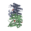 3ts7C 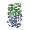 3ucaC 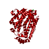 4dhdC 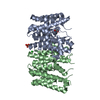 4f62C  4fp4C C: citing same article ( |
|---|---|
| Similar structure data | |
| Other databases |


-Links
 PDBj
PDBj
- Assembly
| Deposited unit | 
| ||||||||
|---|---|---|---|---|---|---|---|---|---|
| 1 | 
| ||||||||
| 2 | 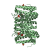
| ||||||||
| 3 | 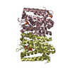
| ||||||||
| Unit cell |
| ||||||||
| Components on special symmetry positions |
|
-Components
| #1: Protein | Mass: 35175.188 Da / Num. of mol.: 4 Source method: isolated from a genetically manipulated source Source: (gene. exp.) Helicobacter pylori (bacteria) / Strain: 26695 / Gene: HP_0929 / Plasmid: pSGX2(BN) / Production host: #2: Chemical | ChemComp-MG /   Mass: 24.305 Da / Num. of mol.: 5 / Source method: obtained synthetically / Formula: Mg Mass: 24.305 Da / Num. of mol.: 5 / Source method: obtained synthetically / Formula: Mg#3: Chemical | ChemComp-DMA /   Mass: 246.092 Da / Num. of mol.: 8 / Source method: obtained synthetically / Formula: C5H12O7P2 Mass: 246.092 Da / Num. of mol.: 8 / Source method: obtained synthetically / Formula: C5H12O7P2#4: Chemical | ChemComp-SO4 /   Mass: 96.063 Da / Num. of mol.: 7 / Source method: obtained synthetically / Formula: SO4 Mass: 96.063 Da / Num. of mol.: 7 / Source method: obtained synthetically / Formula: SO4#5: Water | ChemComp-HOH / |  Mass: 18.015 Da / Num. of mol.: 224 / Source method: isolated from a natural source / Formula: H2O Mass: 18.015 Da / Num. of mol.: 224 / Source method: isolated from a natural source / Formula: H2O |
|---|
-Experimental details
-Experiment
| Experiment | Method: X-RAY DIFFRACTION / Number of used crystals: 1 |
|---|
- Sample preparation
| Crystal | Density Matthews: 4.7 Å3/Da / Density % sol: 73.8 % |
|---|---|
| Crystal grow | Method: vapor diffusion, sitting drop / pH: 7.5 Details: 0.1M HEPES PH 7.5, 2000MM AMMONIUM SULFATE, 10% GLYCEROL, VAPOR DIFFUSION, SITTING DROP, TEMPERATURE 294K |
-Data collection
| Diffraction | Mean temperature: 93 K |
|---|---|
| Diffraction source | Source: SYNCHROTRON / Site: NSLS  / Beamline: X29A / Wavelength: 0.979 / Beamline: X29A / Wavelength: 0.979 |
| Detector | Type: ADSC QUANTUM 315 / Detector: CCD / Date: Jul 15, 2010 / Details: MIRRORS |
| Radiation | Protocol: SINGLE WAVELENGTH / Monochromatic (M) / Laue (L): M / Scattering type: x-ray |
| Radiation wavelength | Wavelength: 0.979 Å / Relative weight: 1 |
| Reflection | Resolution: 2.3→40 Å / Num. obs: 105082 / % possible obs: 100 % / Observed criterion σ(I): -5 / Redundancy: 7.6 % / Biso Wilson estimate: 57.956 Å2 / Rsym value: 0.11 / Net I/σ(I): 8.3 |
| Reflection shell | Resolution: 2.3→2.35 Å / Redundancy: 6.8 % / Mean I/σ(I) obs: 0.6 / % possible all: 100 |
- Processing
| Software |
| ||||||||||||||||||||||||||||||||||||||||||||||||||||||||||||||||||||||||||||||||||||||||||||||||||||||||||||||||||||||||||||||||||||||||||||||||||||||||||||||||||||||||||
|---|---|---|---|---|---|---|---|---|---|---|---|---|---|---|---|---|---|---|---|---|---|---|---|---|---|---|---|---|---|---|---|---|---|---|---|---|---|---|---|---|---|---|---|---|---|---|---|---|---|---|---|---|---|---|---|---|---|---|---|---|---|---|---|---|---|---|---|---|---|---|---|---|---|---|---|---|---|---|---|---|---|---|---|---|---|---|---|---|---|---|---|---|---|---|---|---|---|---|---|---|---|---|---|---|---|---|---|---|---|---|---|---|---|---|---|---|---|---|---|---|---|---|---|---|---|---|---|---|---|---|---|---|---|---|---|---|---|---|---|---|---|---|---|---|---|---|---|---|---|---|---|---|---|---|---|---|---|---|---|---|---|---|---|---|---|---|---|---|---|---|---|
| Refinement | Method to determine structure: SAD / Resolution: 2.4→40 Å / Cor.coef. Fo:Fc: 0.952 / Cor.coef. Fo:Fc free: 0.946 / SU B: 7.57 / SU ML: 0.168 / Cross valid method: THROUGHOUT / ESU R: 0.234 / ESU R Free: 0.198 / Stereochemistry target values: MAXIMUM LIKELIHOOD / Details: HYDROGENS HAVE BEEN ADDED IN THE RIDING POSITIONS
| ||||||||||||||||||||||||||||||||||||||||||||||||||||||||||||||||||||||||||||||||||||||||||||||||||||||||||||||||||||||||||||||||||||||||||||||||||||||||||||||||||||||||||
| Solvent computation | Ion probe radii: 0.8 Å / Shrinkage radii: 0.8 Å / VDW probe radii: 1.2 Å / Solvent model: BABINET MODEL WITH MASK | ||||||||||||||||||||||||||||||||||||||||||||||||||||||||||||||||||||||||||||||||||||||||||||||||||||||||||||||||||||||||||||||||||||||||||||||||||||||||||||||||||||||||||
| Displacement parameters | Biso mean: 63.151 Å2
| ||||||||||||||||||||||||||||||||||||||||||||||||||||||||||||||||||||||||||||||||||||||||||||||||||||||||||||||||||||||||||||||||||||||||||||||||||||||||||||||||||||||||||
| Refinement step | Cycle: LAST / Resolution: 2.4→40 Å
| ||||||||||||||||||||||||||||||||||||||||||||||||||||||||||||||||||||||||||||||||||||||||||||||||||||||||||||||||||||||||||||||||||||||||||||||||||||||||||||||||||||||||||
| Refine LS restraints |
| ||||||||||||||||||||||||||||||||||||||||||||||||||||||||||||||||||||||||||||||||||||||||||||||||||||||||||||||||||||||||||||||||||||||||||||||||||||||||||||||||||||||||||
| LS refinement shell | Resolution: 2.4→2.462 Å / Total num. of bins used: 20
|
