- PDB-4g6c: Crystal structure of beta-hexosaminidase 1 from Burkholderia ceno... -
+
Open data
ID or keywords:
Loading...
-
Basic information
Entry
Database: PDB / ID: 4g6c
Title
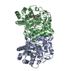
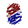
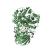
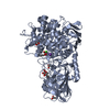
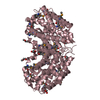
Crystal structure of beta-hexosaminidase 1 from Burkholderia cenocepacia J2315
Components
Beta-hexosaminidase 1
Keywords
HYDROLASE / SSGCID / NIAID / BETA- HEXOSAMINIDASE / Structural Genomics / National Institute of Allergy and Infectious Diseases / Seattle Structural Genomics Center for Infectious Disease
Function / homology
Function and homology information
beta-N-acetylhexosaminidase activity / beta-N-acetylhexosaminidase / peptidoglycan turnover / peptidoglycan biosynthetic process / cell wall organization / regulation of cell shape / carbohydrate metabolic process / cell division / cytoplasm Similarity search - Function
Beta-hexosaminidase, bacterial / : / Glycoside hydrolase, family 3, N-terminal domain / Glycoside hydrolase, family 3, N-terminal / Glycoside hydrolase, family 3, N-terminal domain superfamily / Glycosyl hydrolase family 3 N terminal domain / Glycoside hydrolase superfamily / TIM Barrel / Alpha-Beta Barrel / Alpha Beta Similarity search - Domain/homology
Mass: 18.015 Da / Num. of mol.: 558 / Source method: isolated from a natural source / Formula: H2O
-
Experimental details
-
Experiment
Experiment
Method: X-RAY DIFFRACTION / Number of used crystals: 1
-
Sample preparation
Crystal
Density Matthews: 1.98 Å3/Da / Density % sol: 37.89 %
Crystal grow
Temperature: 289 K / Method: vapor diffusion, sitting drop / pH: 8.5 Details: EBS INTERNAL TRACKING NUMBER 234629B12. : BUCEA.18451.A PW36254 AT 20 MG/ML IN 25 MM HEPES (pH 7.0), 500 mM NaCl, 2 mM DTT, 0.025% sodium azide, 5% glycerol, 0.4 uL x 0.4 uL drop with ...Details: EBS INTERNAL TRACKING NUMBER 234629B12. : BUCEA.18451.A PW36254 AT 20 MG/ML IN 25 MM HEPES (pH 7.0), 500 mM NaCl, 2 mM DTT, 0.025% sodium azide, 5% glycerol, 0.4 uL x 0.4 uL drop with MORPHEUS SCREEN B12: 90 mM Halogens (NaF, NaBr, NaI, 0.1 M Tris/Bicine pH 8.5, 37.5% MPD-PEG1000-PEG3350, VAPOR DIFFUSION, SITTING DROP, temperature 289K
-
Data collection
Diffraction
ID
Mean temperature (K)
Crystal-ID
1
100
1
2
100
1
Diffraction source
Source
Site
Beamline
Type
ID
Wavelength
Wavelength (Å)
SYNCHROTRON
SSRL
BL7-1
1
1.03317
1.03317
ROTATING ANODE
RIGAKU FR-E+ SUPERBRIGHT
2
1.54
1.54
Detector
Type
ID
Detector
Date
ADSC QUANTUM 315r
1
CCD
May 31, 2012
RIGAKU SATURN 944+
2
CCD
May 25, 2012
Radiation
Protocol: SINGLE WAVELENGTH / Monochromatic (M) / Laue (L): M / Scattering type: x-ray
Radiation wavelength
ID
Wavelength (Å)
Relative weight
1
1.03317
1
2
1.54
1
Reflection
Number: 588960 / Rmerge(I) obs: 0.035 / D res high: 1.7 Å / Num. obs: 123462 / % possible obs: 98.9
Diffraction reflection shell
Highest resolution (Å)
Lowest resolution (Å)
Num. obs
% possible obs (%)
ID
Rmerge(I) obs
1.7
1.74
8203
88.8
1
0.18
1.74
1.79
8865
98.6
1
0.164
1.79
1.84
8611
99.1
1
0.139
1.84
1.9
8475
99.3
1
0.12
1.9
1.96
8259
99.8
1
0.096
1.96
2.03
7990
100
1
0.078
2.03
2.11
7642
100
1
0.068
2.11
2.19
7409
100
1
0.057
2.19
2.29
7073
100
1
0.052
2.29
2.4
6784
100
1
0.045
2.4
2.53
6464
100
1
0.041
2.69
2.87
5723
100
1
0.036
2.87
3.1
5330
100
1
0.033
3.1
3.4
4907
100
1
0.029
3.4
3.8
4447
100
1
0.026
3.8
4.39
3910
100
1
0.025
4.39
5.38
3295
100
1
0.024
5.38
7.6
2556
100
1
0.026
Reflection
Resolution: 1.38→99.7 Å / Num. obs: 118123 / % possible obs: 99.7 % / Observed criterion σ(I): -3 / Biso Wilson estimate: 18.888 Å2 / Rmerge(I) obs: 0.047 / Net I/σ(I): 22.51
Reflection shell
Resolution (Å)
Rmerge(I) obs
Mean I/σ(I) obs
Num. measured obs
Num. unique obs
Diffraction-ID
% possible all
1.38-1.42
0.492
3.6
47521
8708
1
99.9
1.42-1.45
0.403
4.42
46622
8508
1
99.9
1.45-1.5
0.332
5.38
45485
8288
1
99.9
1.5-1.54
0.254
6.93
44160
8009
1
99.8
1.54-1.59
0.213
8.36
43075
7787
1
99.8
1.59-1.65
0.167
10.49
41699
7523
1
99.9
1.65-1.71
0.14
12.44
40479
7278
1
99.8
1.71-1.78
0.11
15.2
39029
6991
1
99.8
1.78-1.86
0.087
18.55
37686
6746
1
99.8
1.86-1.95
0.065
23.57
36046
6419
1
99.8
1.95-2.06
0.051
29.06
33954
6072
1
99.8
2.06-2.18
0.043
33.84
32457
5785
1
99.7
2.18-2.33
0.037
38.04
30528
5437
1
99.8
2.33-2.52
0.033
42.75
27996
5029
1
99.8
2.52-2.76
0.031
46.41
25698
4667
1
99.6
2.76-3.09
0.027
50.3
22577
4239
1
99.8
3.09-3.56
0.023
58.26
20782
3726
1
100
3.56-4.36
0.022
62.87
17424
3172
1
99.9
4.36-6.17
0.022
62.64
13369
2471
1
99.9
-
Phasing
Phasing
Method
SAD
molecular replacement
Phasing MR
Model details: Phaser MODE: MR_AUTO
Highest resolution
Lowest resolution
Rotation
2.5 Å
19.7 Å
Translation
2.5 Å
19.7 Å
-
Processing
Software
Name
Version
Classification
NB
XSCALE
datascaling
PHASER
2.3.0
phasing
REFMAC
refinement
PDB_EXTRACT
3.11
dataextraction
XDS
datareduction
Refinement
Method to determine structure: MOLECULAR REPLACEMENT / Resolution: 1.38→99.7 Å / Cor.coef. Fo:Fc: 0.969 / Cor.coef. Fo:Fc free: 0.956 / WRfactor Rfree: 0.1892 / WRfactor Rwork: 0.1668 / Occupancy max: 1 / Occupancy min: 0.5 / FOM work R set: 0.8823 / SU B: 1.968 / SU ML: 0.036 / SU R Cruickshank DPI: 0.0599 / SU Rfree: 0.0608 / Cross valid method: THROUGHOUT / σ(F): 0 / ESU R: 0.065 / ESU R Free: 0.059 / Stereochemistry target values: MAXIMUM LIKELIHOOD Details: HYDROGENS HAVE BEEN USED IF PRESENT IN THE INPUT U VALUES : WITH TLS ADDED
Rfactor
Num. reflection
% reflection
Selection details
Rfree
0.188
5916
5 %
RANDOM
Rwork
0.155
-
-
-
obs
0.157
118094
-
-
Solvent computation
Ion probe radii: 0.8 Å / Shrinkage radii: 0.8 Å / VDW probe radii: 1.2 Å / Solvent model: MASK
In the structure databanks used in Yorodumi, some data are registered as the other names, "COVID-19 virus" and "2019-nCoV". Here are the details of the virus and the list of structure data.
Jan 31, 2019. EMDB accession codes are about to change! (news from PDBe EMDB page)
EMDB accession codes are about to change! (news from PDBe EMDB page)
The allocation of 4 digits for EMDB accession codes will soon come to an end. Whilst these codes will remain in use, new EMDB accession codes will include an additional digit and will expand incrementally as the available range of codes is exhausted. The current 4-digit format prefixed with “EMD-” (i.e. EMD-XXXX) will advance to a 5-digit format (i.e. EMD-XXXXX), and so on. It is currently estimated that the 4-digit codes will be depleted around Spring 2019, at which point the 5-digit format will come into force.
The EM Navigator/Yorodumi systems omit the EMD- prefix.
Related info.:Q: What is EMD? / ID/Accession-code notation in Yorodumi/EM Navigator
Yorodumi is a browser for structure data from EMDB, PDB, SASBDB, etc.
This page is also the successor to EM Navigator detail page, and also detail information page/front-end page for Omokage search.
The word "yorodu" (or yorozu) is an old Japanese word meaning "ten thousand". "mi" (miru) is to see.
Related info.:EMDB / PDB / SASBDB / Comparison of 3 databanks / Yorodumi Search / Aug 31, 2016. New EM Navigator & Yorodumi / Yorodumi Papers / Jmol/JSmol / Function and homology information / Changes in new EM Navigator and Yorodumi
 Movie
Movie Controller

 Yorodumi
 Open data
 Basic information
 Components
 Keywords
 Function and homology information
 Burkholderia cenocepacia (bacteria)
 X-RAY DIFFRACTION /
  Authors
 Citation
 Structure visualization
 Downloads & links
 Other downloads
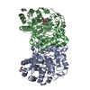
 PDBj
PDBj Assembly
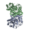
 Mass: 18.015 Da / Num. of mol.: 558 / Source method: isolated from a natural source / Formula: H2O
 Sample preparation
 Processing