 Movie
Movie Controller

[English] 日本語
 Yorodumi
- PDB-6y2n: Crystal structure of ribonucleotide reductase R2 subunit solved b... -
+ Open data

- Basic information
| Entry | Database: PDB / ID: 6y2n | ||||||||||||
|---|---|---|---|---|---|---|---|---|---|---|---|---|---|
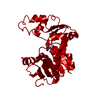
| Title | Crystal structure of ribonucleotide reductase R2 subunit solved by serial synchrotron crystallography | ||||||||||||
 Components Components | Ribonucleoside-diphosphate reductase subunit beta | ||||||||||||
 Keywords Keywords | OXIDOREDUCTASE / RIBONUCLEOTIDE REDUCTASE R2 SUBUNIT / MN/FE COFACTOR / METALLOPROTEIN OXIDOREDUCTASE / FERRITIN-LIKE SUPERFAMILY | ||||||||||||
| Function / homology |  Function and homology information Function and homology informationribonucleoside-diphosphate reductase / ribonucleoside-diphosphate reductase activity, thioredoxin disulfide as acceptor / deoxyribonucleotide biosynthetic process / metal ion binding Similarity search - Function | ||||||||||||
| Biological species |  Saccharopolyspora erythraea (bacteria) Saccharopolyspora erythraea (bacteria) | ||||||||||||
| Method |  X-RAY DIFFRACTION / SYNCHROTRON / FOURIER SYNTHESIS / Resolution: 2.4 Å X-RAY DIFFRACTION / SYNCHROTRON / FOURIER SYNTHESIS / Resolution: 2.4 Å | ||||||||||||
 Authors Authors | Shilova, A. / Lebrette, H. / Aurelius, O. / Hogbom, M. / Mueller, U. | ||||||||||||
| Funding support |  Sweden, 3items Sweden, 3items
| ||||||||||||
 Citation Citation | Journal: J.Synchrotron Radiat. / Year: 2020 Title: Current status and future opportunities for serial crystallography at MAX IV Laboratory. Authors: Shilova, A. / Lebrette, H. / Aurelius, O. / Nan, J. / Welin, M. / Kovacic, R. / Ghosh, S. / Safari, C. / Friel, R.J. / Milas, M. / Matej, Z. / Hogbom, M. / Branden, G. / Kloos, M. / Shoeman, ...Authors: Shilova, A. / Lebrette, H. / Aurelius, O. / Nan, J. / Welin, M. / Kovacic, R. / Ghosh, S. / Safari, C. / Friel, R.J. / Milas, M. / Matej, Z. / Hogbom, M. / Branden, G. / Kloos, M. / Shoeman, R.L. / Doak, B. / Ursby, T. / Hakansson, M. / Logan, D.T. / Mueller, U. | ||||||||||||
| History |
|
- Structure visualization
| Structure viewer | Molecule: MolmilJmol/JSmol |
|---|
- Downloads & links
-Download
| PDBx/mmCIF format | 6y2n.cif.gz | 148.4 KB | Display | PDBx/mmCIF format |
|---|---|---|---|---|
| PDB format | pdb6y2n.ent.gz | 111.2 KB | Display | PDB format |
| PDBx/mmJSON format | 6y2n.json.gz | Tree view | PDBx/mmJSON format | |
| Others |  Other downloads Other downloads |
-Validation report
| Arichive directory | https://data.pdbj.org/pub/pdb/validation_reports/y2/6y2nftp://data.pdbj.org/pub/pdb/validation_reports/y2/6y2n | HTTPS FTP |
|---|
-Related structure data


-Links
 PDBj
PDBj

- Assembly
| Deposited unit | 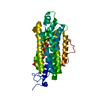
| ||||||||||||
|---|---|---|---|---|---|---|---|---|---|---|---|---|---|
| 1 | 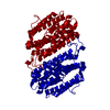
| ||||||||||||
| Unit cell |
| ||||||||||||
| Components on special symmetry positions |
|
-Components
| #1: Protein | Mass: 39608.891 Da / Num. of mol.: 1 Source method: isolated from a genetically manipulated source Source: (gene. exp.) Saccharopolyspora erythraea (strain ATCC 11635 / DSM 40517 / JCM 4748 / NBRC 13426 / NCIMB 8594 / NRRL 2338) (bacteria)Gene: nrdB, SACE_1282, A8924_1677 / Production host: References: UniProt: A4F980, ribonucleoside-diphosphate reductase |
|---|---|
| #2: Chemical | ChemComp-MN3 /   Mass: 54.938 Da / Num. of mol.: 1 / Source method: isolated from a natural source / Formula: Mn Mass: 54.938 Da / Num. of mol.: 1 / Source method: isolated from a natural source / Formula: Mn |
| #3: Chemical | ChemComp-FE /   Mass: 55.845 Da / Num. of mol.: 1 / Source method: obtained synthetically / Formula: Fe Mass: 55.845 Da / Num. of mol.: 1 / Source method: obtained synthetically / Formula: Fe |
| #4: Water | ChemComp-HOH /  Mass: 18.015 Da / Num. of mol.: 23 / Source method: isolated from a natural source / Formula: H2O Mass: 18.015 Da / Num. of mol.: 23 / Source method: isolated from a natural source / Formula: H2O |
| Has ligand of interest | N |
-Experimental details
-Experiment
| Experiment | Method: X-RAY DIFFRACTION / Number of used crystals: 1 |
|---|
- Sample preparation
| Crystal | Density Matthews: 2 Å3/Da / Density % sol: 38.49 % / Description: square bipyramid |
|---|---|
| Crystal grow | Temperature: 294 K / Method: batch mode / pH: 4.5 Details: 16% (w/v) polyethylene glycol 3350, 2% (v/v) tacsimate pH 4.5 |
-Data collection
| Diffraction | Mean temperature: 293 K / Serial crystal experiment: Y |
|---|---|
| Diffraction source | Source: SYNCHROTRON / Site: MAX IV / Beamline: BioMAX / Wavelength: 0.98 Å |
| Detector | Type: DECTRIS EIGER X 16M / Detector: PIXEL / Date: Feb 23, 2019 |
| Radiation | Protocol: SINGLE WAVELENGTH / Monochromatic (M) / Laue (L): M / Scattering type: x-ray |
| Radiation wavelength | Wavelength: 0.98 Å / Relative weight: 1 |
| Reflection | Resolution: 2.4→59.3 Å / Num. obs: 13245 / % possible obs: 98.6 % / Redundancy: 516 % / Biso Wilson estimate: 81.8 Å2 / CC1/2: 0.99 / Net I/σ(I): 6.73 |
| Reflection shell | Resolution: 2.4→2.58 Å / Num. unique obs: 2336 / CC1/2: 0.64 |
| Serial crystallography sample delivery | Description: Silicon nitride membranes / Method: fixed target |
- Processing
| Software |
| ||||||||||||||||||||||||||||||||||||||||||
|---|---|---|---|---|---|---|---|---|---|---|---|---|---|---|---|---|---|---|---|---|---|---|---|---|---|---|---|---|---|---|---|---|---|---|---|---|---|---|---|---|---|---|---|
| Refinement | Method to determine structure: FOURIER SYNTHESIS Starting model: Unpublished Resolution: 2.4→59.3 Å / SU ML: 0.206 / Cross valid method: FREE R-VALUE / σ(F): 0 / Phase error: 25.9869 Stereochemistry target values: GeoStd + Monomer Library + CDL v1.2
| ||||||||||||||||||||||||||||||||||||||||||
| Solvent computation | Shrinkage radii: 0.9 Å / VDW probe radii: 1.11 Å / Solvent model: FLAT BULK SOLVENT MODEL | ||||||||||||||||||||||||||||||||||||||||||
| Displacement parameters | Biso mean: 90.39 Å2 | ||||||||||||||||||||||||||||||||||||||||||
| Refinement step | Cycle: LAST / Resolution: 2.4→59.3 Å
| ||||||||||||||||||||||||||||||||||||||||||
| Refine LS restraints |
| ||||||||||||||||||||||||||||||||||||||||||
| LS refinement shell |
|
