Tripartitemotif-containingprotein54 / Muscle-specific RING finger protein / MuRF / Muscle-specific RING finger protein 3 / MuRF-3 / MuRF3 ...Muscle-specific RING finger protein / MuRF / Muscle-specific RING finger protein 3 / MuRF-3 / MuRF3 / RING finger protein 30
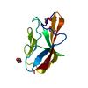
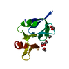
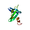
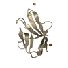
Mass: 5390.276 Da / Num. of mol.: 1 / Fragment: B-Box domain, UNP residues 122-168 Source method: isolated from a genetically manipulated source Source: (gene. exp.) Homo sapiens (human) / Gene: MURF, MURF3, RNF30, TRIM54 / Plasmid: PET28-MHL / Production host: Escherichia coli (E. coli) / Strain (production host): BL21 (DE3) References: UniProt: Q9BYV2, Ligases; Forming carbon-nitrogen bonds; Acid-amino-acid ligases (peptide synthases)
Mass: 18.015 Da / Num. of mol.: 32 / Source method: isolated from a natural source / Formula: H2O
Sequence details
THE SEQUENCE PUT IN FOR CRYSTALLIZATION IS ...THE SEQUENCE PUT IN FOR CRYSTALLIZATION IS MHHHHHHSSGRENLYFQGQESSRPLHSKAEQHLMCEEHEEEKINIYCLSCEVPTCSLCKVFGAHKDCEVAPLPTIYKRQK AND IN SITU PROTEOLYSIS HAS BEEN CARRIED OUT DURING CRYSTALLIZATION
-
Experimental details
-
Experiment
Experiment
Method: X-RAY DIFFRACTION / Number of used crystals: 1
-
Sample preparation
Crystal
Density Matthews: 2.62 Å3/Da / Density % sol: 52.97 %
Resolution: 2.15→29.823 Å / Cor.coef. Fo:Fc: 0.952 / Cor.coef. Fo:Fc free: 0.897 / SU B: 7.524 / SU ML: 0.109 / Cross valid method: THROUGHOUT / ESU R Free: 0.194 / Stereochemistry target values: MAXIMUM LIKELIHOOD / Details: HYDROGENS HAVE BEEN ADDED IN THE RIDING POSITIONS
Rfactor
Num. reflection
% reflection
Selection details
Rfree
0.25343
148
4.3 %
RANDOM
Rwork
0.18198
-
-
-
obs
0.18518
3297
99.11 %
-
all
-
3560
-
-
Solvent computation
Ion probe radii: 0.8 Å / Shrinkage radii: 0.8 Å / VDW probe radii: 1.4 Å / Solvent model: MASK
Displacement parameters
Biso mean: 25.228 Å2
Baniso -1
Baniso -2
Baniso -3
1-
0.94 Å2
0.47 Å2
0 Å2
2-
-
0.94 Å2
0 Å2
3-
-
-
-1.4 Å2
Refinement step
Cycle: LAST / Resolution: 2.15→29.823 Å
Protein
Nucleic acid
Ligand
Solvent
Total
Num. atoms
364
0
2
32
398
Refine LS restraints
Refine-ID
Type
Dev ideal
Dev ideal target
Number
X-RAY DIFFRACTION
r_bond_refined_d
0.011
0.022
387
X-RAY DIFFRACTION
r_angle_refined_deg
1.417
1.987
520
X-RAY DIFFRACTION
r_dihedral_angle_1_deg
6.805
5
48
X-RAY DIFFRACTION
r_dihedral_angle_2_deg
36.411
25.714
14
X-RAY DIFFRACTION
r_dihedral_angle_3_deg
13.624
15
68
X-RAY DIFFRACTION
r_chiral_restr
0.087
0.2
59
X-RAY DIFFRACTION
r_gen_planes_refined
0.006
0.021
277
X-RAY DIFFRACTION
r_mcbond_it
0.454
1.5
242
X-RAY DIFFRACTION
r_mcangle_it
0.875
2
395
X-RAY DIFFRACTION
r_scbond_it
2.843
3
145
X-RAY DIFFRACTION
r_scangle_it
3.584
4.5
125
LS refinement shell
Resolution: 2.151→2.206 Å / Total num. of bins used: 20
Rfactor
Num. reflection
% reflection
Rfree
0.264
6
-
Rwork
0.182
230
-
obs
-
-
99.16 %
Refinement TLS params.
Method: refined / Refine-ID: X-RAY DIFFRACTION
ID
L11 (°2)
L12 (°2)
L13 (°2)
L22 (°2)
L23 (°2)
L33 (°2)
S11 (Å °)
S12 (Å °)
S13 (Å °)
S21 (Å °)
S22 (Å °)
S23 (Å °)
S31 (Å °)
S32 (Å °)
S33 (Å °)
T11 (Å2)
T12 (Å2)
T13 (Å2)
T22 (Å2)
T23 (Å2)
T33 (Å2)
Origin x (Å)
Origin y (Å)
Origin z (Å)
1
29.9619
-10.8967
10.7982
15.0728
-5.0377
22.7087
0.5157
-0.794
-1.2503
-0.1348
0.4258
0.3911
1.3049
-0.3697
-0.9415
0.1204
-0.0754
-0.0739
0.1004
0.0638
0.0771
20.8677
8.5902
9.6691
2
16.3714
-9.1763
-1.6635
13.8175
-3.6247
3.3621
0.2474
0.5132
-1.013
-0.4851
-0.1549
0.5592
0.3107
-0.1847
-0.0924
0.1079
-0.0436
-0.0859
0.2059
-0.0025
0.13
16.0162
11.4292
3.5639
3
7.4752
-0.5072
-0.7907
6.66
-0.0013
4.4855
0.05
-0.5503
-0.4324
-0.4763
0.0266
-0.3333
-0.1158
-0.0227
-0.0766
0.0408
-0.022
0.0251
0.1633
0.0413
0.0488
25.2163
16.582
9.152
4
3.5023
-2.1846
1.1651
8.2582
-4.9403
2.9627
-0.0723
-1.1021
0.157
0.4282
-0.0395
-0.23
-0.2625
0.0641
0.1119
0.0864
-0.0286
-0.0096
0.5044
-0.0894
0.1146
23.1805
23.6783
14.7455
5
6.3319
-0.8829
5.1653
8.6208
-3.5893
8.2642
0.1693
-0.2133
-0.0414
-0.0381
-0.031
0.0595
0.1957
-0.036
-0.1383
0.0329
-0.019
0.0086
0.1273
0.0094
0.0251
20.8758
19.225
4.3376
6
3.5157
-0.7929
0.6439
8.9338
12.4115
19.8874
-0.1033
-0.1269
-0.0501
-0.1731
-0.0916
0.2307
-0.4523
-0.7319
0.1949
0.0207
0.0538
0.0014
0.179
0.0009
0.1007
17.5702
23.6628
2.9841
7
35.5361
-13.0259
-1.7172
13.5571
5.2098
2.4727
0.3835
-0.2375
2.3241
-0.1558
-0.4355
-0.5509
-0.0235
-0.2555
0.052
0.0627
-0.0014
0.0299
0.1885
-0.0456
0.1638
25.7533
26.162
7.6566
8
25.9028
4.3338
-9.1789
5.374
-3.7254
18.2267
0.0854
0.1586
-0.9727
-0.3027
-0.332
-0.5637
1.1365
-0.0379
0.2466
0.1216
-0.0242
-0.0184
0.1299
0.0628
0.0955
34.025
13.8303
7.2466
Refinement TLS group
ID
Refine-ID
Refine TLS-ID
Auth asym-ID
Auth seq-ID
1
X-RAY DIFFRACTION
1
A
122 - 127
2
X-RAY DIFFRACTION
2
A
128 - 132
3
X-RAY DIFFRACTION
3
A
133 - 138
4
X-RAY DIFFRACTION
4
A
139 - 144
5
X-RAY DIFFRACTION
5
A
145 - 150
6
X-RAY DIFFRACTION
6
A
151 - 156
7
X-RAY DIFFRACTION
7
A
157 - 162
8
X-RAY DIFFRACTION
8
A
163 - 168
+
About Yorodumi
-
News
-
Feb 9, 2022. New format data for meta-information of EMDB entries
New format data for meta-information of EMDB entries
Version 3 of the EMDB header file is now the official format.
The previous official version 1.9 will be removed from the archive.
In the structure databanks used in Yorodumi, some data are registered as the other names, "COVID-19 virus" and "2019-nCoV". Here are the details of the virus and the list of structure data.
Jan 31, 2019. EMDB accession codes are about to change! (news from PDBe EMDB page)
EMDB accession codes are about to change! (news from PDBe EMDB page)
The allocation of 4 digits for EMDB accession codes will soon come to an end. Whilst these codes will remain in use, new EMDB accession codes will include an additional digit and will expand incrementally as the available range of codes is exhausted. The current 4-digit format prefixed with “EMD-” (i.e. EMD-XXXX) will advance to a 5-digit format (i.e. EMD-XXXXX), and so on. It is currently estimated that the 4-digit codes will be depleted around Spring 2019, at which point the 5-digit format will come into force.
The EM Navigator/Yorodumi systems omit the EMD- prefix.
Related info.:Q: What is EMD? / ID/Accession-code notation in Yorodumi/EM Navigator
Yorodumi is a browser for structure data from EMDB, PDB, SASBDB, etc.
This page is also the successor to EM Navigator detail page, and also detail information page/front-end page for Omokage search.
The word "yorodu" (or yorozu) is an old Japanese word meaning "ten thousand". "mi" (miru) is to see.
Related info.:EMDB / PDB / SASBDB / Comparison of 3 databanks / Yorodumi Search / Aug 31, 2016. New EM Navigator & Yorodumi / Yorodumi Papers / Jmol/JSmol / Function and homology information / Changes in new EM Navigator and Yorodumi
 Movie
Movie Controller


 Open data
 Basic information
 Components
 Keywords
 Function and homology information
 Homo sapiens (human)
 X-RAY DIFFRACTION /
  Authors
 Citation
 Structure visualization
 Downloads & links
 Other downloads
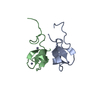


 PDBj
PDBj

 Assembly
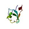


 Mass: 65.409 Da / Num. of mol.: 2 / Source method: obtained synthetically / Formula: Zn
 Mass: 18.015 Da / Num. of mol.: 32 / Source method: isolated from a natural source / Formula: H2O
 Sample preparation
 / Beamline: 19-ID / Wavelength: 0.97951 / Wavelength: 0.97951 Å
 Processing