 Movie
Movie Controller

[English] 日本語
 Yorodumi
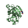
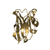
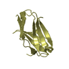
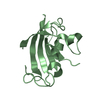
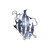
- PDB-1avs: X-RAY CRYSTALLOGRAPHIC STUDY OF CALCIUM-SATURATED N-TERMINAL DOMA... -
+ Open data

- Basic information
| Entry | Database: PDB / ID: 1avs | ||||||
|---|---|---|---|---|---|---|---|
| Title | X-RAY CRYSTALLOGRAPHIC STUDY OF CALCIUM-SATURATED N-TERMINAL DOMAIN OF TROPONIN C | ||||||
 Components Components | TROPONIN C | ||||||
 Keywords Keywords | MUSCLE CONTRACTION / CALCIUM-ACTIVATED / TROPONIN / E-F HAND CALCIUM-BINDING PROTEIN | ||||||
| Function / homology |  Function and homology information Function and homology informationStriated Muscle Contraction / troponin complex / skeletal muscle contraction / calcium ion binding Similarity search - Function | ||||||
| Biological species |  | ||||||
| Method |  X-RAY DIFFRACTION / SYNCHROTRON / MOLECULAR REPLACEMENT, MIR / Resolution: 1.75 Å X-RAY DIFFRACTION / SYNCHROTRON / MOLECULAR REPLACEMENT, MIR / Resolution: 1.75 Å | ||||||
 Authors Authors | Strynadka, N.C.J. / James, M.N.G. | ||||||
 Citation Citation | Journal: J.Mol.Biol. / Year: 1997 Title: Structural details of a calcium-induced molecular switch: X-ray crystallographic analysis of the calcium-saturated N-terminal domain of troponin C at 1.75 A resolution. Authors: Strynadka, N.C. / Cherney, M. / Sielecki, A.R. / Li, M.X. / Smillie, L.B. / James, M.N. | ||||||
| History |
|
- Structure visualization
| Structure viewer | Molecule: MolmilJmol/JSmol |
|---|
- Downloads & links
-Download
| PDBx/mmCIF format | 1avs.cif.gz | 47.1 KB | Display | PDBx/mmCIF format |
|---|---|---|---|---|
| PDB format | pdb1avs.ent.gz | 33 KB | Display | PDB format |
| PDBx/mmJSON format | 1avs.json.gz | Tree view | PDBx/mmJSON format | |
| Others |  Other downloads Other downloads |
-Validation report
| Arichive directory | https://data.pdbj.org/pub/pdb/validation_reports/av/1avsftp://data.pdbj.org/pub/pdb/validation_reports/av/1avs | HTTPS FTP |
|---|
-Related structure data
| Related structure data |  4tncS S: Starting model for refinement |
|---|---|
| Similar structure data |


-Links
 PDBj
PDBj- Assembly
| Deposited unit | 
| ||||||||
|---|---|---|---|---|---|---|---|---|---|
| 1 | 
| ||||||||
| 2 | 
| ||||||||
| Unit cell |
| ||||||||
| Noncrystallographic symmetry (NCS) | NCS oper: (Code: given Matrix: (0.694896, -0.010889, 0.719028), Vector: |
-Components
| #1: Protein | Mass: 9984.085 Da / Num. of mol.: 2 / Fragment: N-TERMINAL DOMAIN, RESIDUES 1 - 90 Source method: isolated from a genetically manipulated source Source: (gene. exp.)  #2: Chemical | ChemComp-CA /   Mass: 40.078 Da / Num. of mol.: 4 / Source method: obtained synthetically / Formula: Ca Mass: 40.078 Da / Num. of mol.: 4 / Source method: obtained synthetically / Formula: Ca#3: Water | ChemComp-HOH / |  Mass: 18.015 Da / Num. of mol.: 93 / Source method: isolated from a natural source / Formula: H2O Mass: 18.015 Da / Num. of mol.: 93 / Source method: isolated from a natural source / Formula: H2O |
|---|
-Experimental details
-Experiment
| Experiment | Method: X-RAY DIFFRACTION / Number of used crystals: 1 |
|---|
- Sample preparation
| Crystal | Density Matthews: 2.4 Å3/Da / Density % sol: 51 % | ||||||||||||||||||||||||||||||||||||
|---|---|---|---|---|---|---|---|---|---|---|---|---|---|---|---|---|---|---|---|---|---|---|---|---|---|---|---|---|---|---|---|---|---|---|---|---|---|
| Crystal grow | pH: 7.5 Details: 72 % AMMONIUM SULFATE 100 MM TRIS 2 % MPD, 5 MM CALCIUM CHLORIDE PH 7.5 | ||||||||||||||||||||||||||||||||||||
| Crystal grow | *PLUS Method: vapor diffusion, hanging dropDetails: drop solution was mixed with an equal volume of reservoir solution | ||||||||||||||||||||||||||||||||||||
| Components of the solutions | *PLUS
|
-Data collection
| Diffraction | Mean temperature: 287 K |
|---|---|
| Diffraction source | Source: SYNCHROTRON / Site: EMBL/DESY, HAMBURG  / Beamline: X11 / Wavelength: 1.009 / Beamline: X11 / Wavelength: 1.009 |
| Detector | Type: MARRESEARCH / Detector: IMAGE PLATE / Date: Nov 1, 1995 / Details: NO MIRRORS |
| Radiation | Monochromator: NI FILTER / Monochromatic (M) / Laue (L): M / Scattering type: x-ray |
| Radiation wavelength | Wavelength: 1.009 Å / Relative weight: 1 |
| Reflection | Resolution: 1.75→20 Å / Num. obs: 18010 / % possible obs: 82 % / Observed criterion σ(I): 0 / Redundancy: 4.3 % / Biso Wilson estimate: 30.4 Å2 / Rmerge(I) obs: 0.052 / Rsym value: 0.104 / Net I/σ(I): 15 |
| Reflection shell | Resolution: 1.75→2 Å / Redundancy: 2 % / Rmerge(I) obs: 0.05 / Mean I/σ(I) obs: 3 / Rsym value: 0.28 / % possible all: 82 |
| Reflection | *PLUS Num. measured all: 85689 |
| Reflection shell | *PLUS % possible obs: 82 % |
- Processing
| Software |
| ||||||||||||||||||||||||||||||||||||||||||||||||||
|---|---|---|---|---|---|---|---|---|---|---|---|---|---|---|---|---|---|---|---|---|---|---|---|---|---|---|---|---|---|---|---|---|---|---|---|---|---|---|---|---|---|---|---|---|---|---|---|---|---|---|---|
| Refinement | Method to determine structure: MOLECULAR REPLACEMENT, MIR Starting model: PDB ENTRY 4TNC Resolution: 1.75→20 Å / Isotropic thermal model: TNT BCORREL V1.0 / σ(F): 0 / Stereochemistry target values: TNT PROTGEO
| ||||||||||||||||||||||||||||||||||||||||||||||||||
| Solvent computation | Solvent model: BABINET SCALING / Bsol: 300 Å2 / ksol: 0.75 e/Å3 | ||||||||||||||||||||||||||||||||||||||||||||||||||
| Refinement step | Cycle: LAST / Resolution: 1.75→20 Å
| ||||||||||||||||||||||||||||||||||||||||||||||||||
| Refine LS restraints |
| ||||||||||||||||||||||||||||||||||||||||||||||||||
| Software | *PLUS Name: TNT / Version: 5D / Classification: refinement | ||||||||||||||||||||||||||||||||||||||||||||||||||
| Refinement | *PLUS Rfactor obs: 0.214 / Rfactor Rfree: 0.251 | ||||||||||||||||||||||||||||||||||||||||||||||||||
| Solvent computation | *PLUS | ||||||||||||||||||||||||||||||||||||||||||||||||||
| Displacement parameters | *PLUS | ||||||||||||||||||||||||||||||||||||||||||||||||||
| Refine LS restraints | *PLUS
|
