 Movie
Movie Controller

[English] 日本語
 Yorodumi
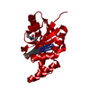
- PDB-3pxu: Crystal structure of phosphopantetheine adenylyltransferase from ... -
+ Open data

- Basic information
| Entry | Database: PDB / ID: 3pxu | |||||||||
|---|---|---|---|---|---|---|---|---|---|---|
| Title | Crystal structure of phosphopantetheine adenylyltransferase from Burkholderia pseudomallei bound to dephospho-coenzyme A | |||||||||
 Components Components | Phosphopantetheine adenylyltransferase | |||||||||
 Keywords Keywords | TRANSFERASE / Structural Genomics / Seattle Structural Genomics Center for Infectious Disease / SSGCID / HUMAN AND ANIMAL PATHOGEN / MELIOIDOSIS / ATP-BINDING / COENZYME A BIOSYNTHESIS / NUCLEOTIDE-BINDING / NUCLEOTIDYLTRANSFERASE / PPAT / pantetheine-phosphate adenylyltransferase | |||||||||
| Function / homology |  Function and homology information Function and homology informationpantetheine-phosphate adenylyltransferase / pantetheine-phosphate adenylyltransferase activity / coenzyme A biosynthetic process / ATP binding / cytoplasm Similarity search - Function | |||||||||
| Biological species |  Burkholderia pseudomallei (bacteria) Burkholderia pseudomallei (bacteria) | |||||||||
| Method |  X-RAY DIFFRACTION / MOLECULAR REPLACEMENT / molecular replacement / Resolution: 2.1 Å X-RAY DIFFRACTION / MOLECULAR REPLACEMENT / molecular replacement / Resolution: 2.1 Å | |||||||||
 Authors Authors | Seattle Structural Genomics Center for Infectious Disease (SSGCID) | |||||||||
 Citation Citation | Journal: Acta Crystallogr.,Sect.F / Year: 2011 Title: Structures of phosphopantetheine adenylyltransferase from Burkholderia pseudomallei. Authors: Edwards, T.E. / Leibly, D.J. / Bhandari, J. / Statnekov, J.B. / Phan, I. / Dieterich, S.H. / Abendroth, J. / Staker, B.L. / Van Voorhis, W.C. / Myler, P.J. / Stewart, L.J. #1: Journal: Plos One / Year: 2013Title: Combining functional and structural genomics to sample the essential Burkholderia structome. Authors: Baugh, L. / Gallagher, L.A. / Patrapuvich, R. / Clifton, M.C. / Gardberg, A.S. / Edwards, T.E. / Armour, B. / Begley, D.W. / Dieterich, S.H. / Dranow, D.M. / Abendroth, J. / Fairman, J.W. / ...Authors: Baugh, L. / Gallagher, L.A. / Patrapuvich, R. / Clifton, M.C. / Gardberg, A.S. / Edwards, T.E. / Armour, B. / Begley, D.W. / Dieterich, S.H. / Dranow, D.M. / Abendroth, J. / Fairman, J.W. / Fox, D. / Staker, B.L. / Phan, I. / Gillespie, A. / Choi, R. / Nakazawa-Hewitt, S. / Nguyen, M.T. / Napuli, A. / Barrett, L. / Buchko, G.W. / Stacy, R. / Myler, P.J. / Stewart, L.J. / Manoil, C. / Van Voorhis, W.C. | |||||||||
| History |
|
- Structure visualization
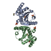
| Structure viewer | Molecule: MolmilJmol/JSmol |
|---|
- Downloads & links
-Download
| PDBx/mmCIF format | 3pxu.cif.gz | 84.3 KB | Display | PDBx/mmCIF format |
|---|---|---|---|---|
| PDB format | pdb3pxu.ent.gz | 62.6 KB | Display | PDB format |
| PDBx/mmJSON format | 3pxu.json.gz | Tree view | PDBx/mmJSON format | |
| Others |  Other downloads Other downloads |
-Validation report
| Arichive directory | https://data.pdbj.org/pub/pdb/validation_reports/px/3pxuftp://data.pdbj.org/pub/pdb/validation_reports/px/3pxu | HTTPS FTP |
|---|
-Related structure data
| Related structure data |  3k9wC  1b6tS C: citing same article ( S: Starting model for refinement |
|---|---|
| Similar structure data | |
| Other databases |


-Links
 PDBj
PDBj- Assembly
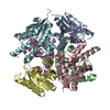
| Deposited unit | 
| ||||||||
|---|---|---|---|---|---|---|---|---|---|
| 1 | x 6
| ||||||||
| 2 | x 24
| ||||||||
| Unit cell |
| ||||||||
| Components on special symmetry positions |
|
-Components
| #1: Protein | Mass: 18849.650 Da / Num. of mol.: 1 Source method: isolated from a genetically manipulated source Source: (gene. exp.) Burkholderia pseudomallei (bacteria) / Strain: 1710b / Gene: BURPS1710b_0748, coaD, coaD BURPS1710B_0748 / Production host: References: UniProt: Q3JW91, pantetheine-phosphate adenylyltransferase | ||||
|---|---|---|---|---|---|
| #2: Chemical | ChemComp-COD / 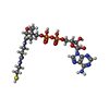  Mass: 687.554 Da / Num. of mol.: 1 / Source method: obtained synthetically / Formula: C21H35N7O13P2S Mass: 687.554 Da / Num. of mol.: 1 / Source method: obtained synthetically / Formula: C21H35N7O13P2S | ||||
| #3: Chemical |   Mass: 92.094 Da / Num. of mol.: 2 / Source method: obtained synthetically / Formula: C3H8O3 Mass: 92.094 Da / Num. of mol.: 2 / Source method: obtained synthetically / Formula: C3H8O3#4: Chemical |   Mass: 96.063 Da / Num. of mol.: 2 / Source method: obtained synthetically / Formula: SO4 Mass: 96.063 Da / Num. of mol.: 2 / Source method: obtained synthetically / Formula: SO4#5: Water | ChemComp-HOH / |  Mass: 18.015 Da / Num. of mol.: 89 / Source method: isolated from a natural source / Formula: H2O Mass: 18.015 Da / Num. of mol.: 89 / Source method: isolated from a natural source / Formula: H2O |
-Experimental details
-Experiment
| Experiment | Method: X-RAY DIFFRACTION / Number of used crystals: 1 |
|---|
- Sample preparation
| Crystal | Density Matthews: 2.67 Å3/Da / Density % sol: 53.91 % |
|---|---|
| Crystal grow | Temperature: 298 K / Method: vapor diffusion, sitting drop / pH: 7 Details: Hampton Crystal Screen condition c8, 2.0 M ammonium sulfate, 5.5 mg/mL protein with expression tag removed, crystal tracking ID 203611c8, pH 7.0, VAPOR DIFFUSION, SITTING DROP, temperature 289K, temperature 298K |
-Data collection
| Diffraction | Mean temperature: 100 K | |||||||||||||||||||||||||||||||||||||||||||||||||||||||||||||||||||||||||||||
|---|---|---|---|---|---|---|---|---|---|---|---|---|---|---|---|---|---|---|---|---|---|---|---|---|---|---|---|---|---|---|---|---|---|---|---|---|---|---|---|---|---|---|---|---|---|---|---|---|---|---|---|---|---|---|---|---|---|---|---|---|---|---|---|---|---|---|---|---|---|---|---|---|---|---|---|---|---|---|
| Diffraction source | Source: ROTATING ANODE / Type: RIGAKU FR-E+ SUPERBRIGHT / Wavelength: 1.5418 Å | |||||||||||||||||||||||||||||||||||||||||||||||||||||||||||||||||||||||||||||
| Detector | Type: RIGAKU SATURN 944+ / Detector: CCD / Date: Aug 4, 2009 | |||||||||||||||||||||||||||||||||||||||||||||||||||||||||||||||||||||||||||||
| Radiation | Protocol: SINGLE WAVELENGTH / Monochromatic (M) / Laue (L): M / Scattering type: x-ray | |||||||||||||||||||||||||||||||||||||||||||||||||||||||||||||||||||||||||||||
| Radiation wavelength | Wavelength: 1.5418 Å / Relative weight: 1 | |||||||||||||||||||||||||||||||||||||||||||||||||||||||||||||||||||||||||||||
| Reflection | Resolution: 2.05→50 Å / Num. obs: 13128 / % possible obs: 99 % / Redundancy: 23.2 % / Rmerge(I) obs: 0.087 / Χ2: 1.097 / Net I/σ(I): 44.5 | |||||||||||||||||||||||||||||||||||||||||||||||||||||||||||||||||||||||||||||
| Reflection shell | Diffraction-ID: 1
|
-Phasing
| Phasing | Method: molecular replacement | |||||||||
|---|---|---|---|---|---|---|---|---|---|---|
| Phasing MR | Rfactor: 56.03 / Model details: Phaser MODE: MR_AUTO
|
- Processing
| Software |
| |||||||||||||||||||||||||||||||||||||||||||||||||||||||||||||||||
|---|---|---|---|---|---|---|---|---|---|---|---|---|---|---|---|---|---|---|---|---|---|---|---|---|---|---|---|---|---|---|---|---|---|---|---|---|---|---|---|---|---|---|---|---|---|---|---|---|---|---|---|---|---|---|---|---|---|---|---|---|---|---|---|---|---|---|
| Refinement | Method to determine structure: MOLECULAR REPLACEMENT Starting model: 1B6T molecule A, protein only Resolution: 2.1→28.6 Å / Cor.coef. Fo:Fc: 0.944 / Cor.coef. Fo:Fc free: 0.933 / WRfactor Rfree: 0.2172 / WRfactor Rwork: 0.1847 / Occupancy max: 1 / Occupancy min: 0.5 / FOM work R set: 0.8339 / SU B: 9.406 / SU ML: 0.114 / SU R Cruickshank DPI: 0.2065 / SU Rfree: 0.1801 / Cross valid method: THROUGHOUT / σ(F): 0 / ESU R Free: 0.18 / Stereochemistry target values: MAXIMUM LIKELIHOOD / Details: HYDROGENS HAVE BEEN ADDED IN THE RIDING POSITIONS.
| |||||||||||||||||||||||||||||||||||||||||||||||||||||||||||||||||
| Solvent computation | Ion probe radii: 0.8 Å / Shrinkage radii: 0.8 Å / VDW probe radii: 1.4 Å / Solvent model: MASK | |||||||||||||||||||||||||||||||||||||||||||||||||||||||||||||||||
| Displacement parameters | Biso max: 76.24 Å2 / Biso mean: 29.9984 Å2 / Biso min: 12.43 Å2 | |||||||||||||||||||||||||||||||||||||||||||||||||||||||||||||||||
| Refinement step | Cycle: LAST / Resolution: 2.1→28.6 Å
| |||||||||||||||||||||||||||||||||||||||||||||||||||||||||||||||||
| Refine LS restraints |
| |||||||||||||||||||||||||||||||||||||||||||||||||||||||||||||||||
| LS refinement shell | Resolution: 2.101→2.155 Å / Total num. of bins used: 20
| |||||||||||||||||||||||||||||||||||||||||||||||||||||||||||||||||
| Refinement TLS params. | Method: refined / Origin x: -20.3175 Å / Origin y: 40.5112 Å / Origin z: -14.7962 Å
|
