 Movie
Movie Controller

[English] 日本語
 Yorodumi
- PDB-3nbk: Phosphopantetheine Adenylyltransferase from Mycobacterium tubercu... -
+ Open data

- Basic information
| Entry | Database: PDB / ID: 3nbk | ||||||
|---|---|---|---|---|---|---|---|
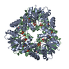
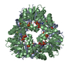
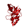
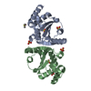
| Title | Phosphopantetheine Adenylyltransferase from Mycobacterium tuberculosis in complex with 4'-phosphopantetheine | ||||||
 Components Components | Phosphopantetheine adenylyltransferase | ||||||
 Keywords Keywords | TRANSFERASE / PPAT / PhP / pantetheine / tuberculosis / phosphopantetheine adenylyltransferase | ||||||
| Function / homology |  Function and homology information Function and homology informationpantetheine-phosphate adenylyltransferase / pantetheine-phosphate adenylyltransferase activity / coenzyme A biosynthetic process / protein hexamerization / ATP binding / cytoplasm Similarity search - Function | ||||||
| Biological species |   Mycobacterium tuberculosis (bacteria) Mycobacterium tuberculosis (bacteria) | ||||||
| Method |  X-RAY DIFFRACTION / SYNCHROTRON / MOLECULAR REPLACEMENT / Resolution: 1.58 Å X-RAY DIFFRACTION / SYNCHROTRON / MOLECULAR REPLACEMENT / Resolution: 1.58 Å | ||||||
 Authors Authors | Wubben, T.J. / Mesecar, A.D. | ||||||
 Citation Citation | Journal: J.Mol.Biol. / Year: 2010 Title: Kinetic, thermodynamic, and structural insight into the mechanism of phosphopantetheine adenylyltransferase from Mycobacterium tuberculosis. Authors: Wubben, T.J. / Mesecar, A.D. | ||||||
| History |
|
- Structure visualization
| Structure viewer | Molecule: MolmilJmol/JSmol |
|---|
- Downloads & links
-Download
| PDBx/mmCIF format | 3nbk.cif.gz | 156.7 KB | Display | PDBx/mmCIF format |
|---|---|---|---|---|
| PDB format | pdb3nbk.ent.gz | 122.9 KB | Display | PDB format |
| PDBx/mmJSON format | 3nbk.json.gz | Tree view | PDBx/mmJSON format | |
| Others |  Other downloads Other downloads |
-Validation report
| Arichive directory | https://data.pdbj.org/pub/pdb/validation_reports/nb/3nbkftp://data.pdbj.org/pub/pdb/validation_reports/nb/3nbk | HTTPS FTP |
|---|
-Related structure data
| Related structure data |  3nbaC  1tfuS C: citing same article ( S: Starting model for refinement |
|---|---|
| Similar structure data |


-Links
 PDBj
PDBj- Assembly
| Deposited unit | 
| ||||||||
|---|---|---|---|---|---|---|---|---|---|
| 1 | 
| ||||||||
| 2 | 
| ||||||||
| Unit cell |
|
-Components
| #1: Protein | Mass: 19331.111 Da / Num. of mol.: 4 Source method: isolated from a genetically manipulated source Source: (gene. exp.) Mycobacterium tuberculosis (bacteria) / Gene: coaD, kdtB, MT3043, MTCY349.22, Rv2965c, u0002e / Plasmid: pET15b / Production host: References: UniProt: P0A530, UniProt: P9WPA5*PLUS, pantetheine-phosphate adenylyltransferase #2: Chemical | ChemComp-PNS /   Mass: 358.348 Da / Num. of mol.: 4 / Source method: obtained synthetically / Formula: C11H23N2O7PS Mass: 358.348 Da / Num. of mol.: 4 / Source method: obtained synthetically / Formula: C11H23N2O7PS#3: Chemical |   Mass: 58.693 Da / Num. of mol.: 2 / Source method: obtained synthetically / Formula: Ni Mass: 58.693 Da / Num. of mol.: 2 / Source method: obtained synthetically / Formula: Ni#4: Water | ChemComp-HOH / |  Mass: 18.015 Da / Num. of mol.: 775 / Source method: isolated from a natural source / Formula: H2O Mass: 18.015 Da / Num. of mol.: 775 / Source method: isolated from a natural source / Formula: H2O |
|---|
-Experimental details
-Experiment
| Experiment | Method: X-RAY DIFFRACTION / Number of used crystals: 1 |
|---|
- Sample preparation
| Crystal | Density Matthews: 3.51 Å3/Da / Density % sol: 64.92 % |
|---|---|
| Crystal grow | Temperature: 294 K / Method: vapor diffusion, hanging drop / pH: 7 Details: 0.05 M Hepes pH 7 and 2.5 v/v % isopropanol, VAPOR DIFFUSION, HANGING DROP, temperature 294K |
-Data collection
| Diffraction | Mean temperature: 77 K |
|---|---|
| Diffraction source | Source: SYNCHROTRON / Site: APS  / Beamline: 22-BM / Wavelength: 1 Å / Beamline: 22-BM / Wavelength: 1 Å |
| Detector | Type: MARMOSAIC 225 mm CCD / Detector: CCD / Date: Aug 13, 2009 |
| Radiation | Monochromator: double-crystal Si (111) sagittal focusing, vertical focusing mirror Protocol: SINGLE WAVELENGTH / Monochromatic (M) / Laue (L): M / Scattering type: x-ray |
| Radiation wavelength | Wavelength: 1 Å / Relative weight: 1 |
| Reflection | Resolution: 1.58→135.21 Å / Num. all: 141372 / Num. obs: 141259 / % possible obs: 99.9 % / Observed criterion σ(I): 2 / Redundancy: 10.5 % / Biso Wilson estimate: 23.3 Å2 / Rmerge(I) obs: 0.094 / Net I/σ(I): 32.9 |
| Reflection shell | Resolution: 1.58→1.61 Å / Redundancy: 8.6 % / Rmerge(I) obs: 0.666 / Mean I/σ(I) obs: 3.4 / Num. unique all: 7008 / % possible all: 99.9 |
- Processing
| Software |
| |||||||||||||||||||||||||||||||||||||||||||||||||||||||||||||||||
|---|---|---|---|---|---|---|---|---|---|---|---|---|---|---|---|---|---|---|---|---|---|---|---|---|---|---|---|---|---|---|---|---|---|---|---|---|---|---|---|---|---|---|---|---|---|---|---|---|---|---|---|---|---|---|---|---|---|---|---|---|---|---|---|---|---|---|
| Refinement | Method to determine structure: MOLECULAR REPLACEMENT Starting model: PDB entry 1TFU Resolution: 1.58→135.21 Å / Cor.coef. Fo:Fc: 0.958 / Cor.coef. Fo:Fc free: 0.952 / SU B: 1.081 / SU ML: 0.039 / Cross valid method: THROUGHOUT / ESU R Free: 0.066 / Stereochemistry target values: MAXIMUM LIKELIHOOD / Details: HYDROGENS HAVE BEEN ADDED IN THE RIDING POSITIONS
| |||||||||||||||||||||||||||||||||||||||||||||||||||||||||||||||||
| Solvent computation | Ion probe radii: 0.8 Å / Shrinkage radii: 0.8 Å / VDW probe radii: 1.4 Å / Solvent model: MASK | |||||||||||||||||||||||||||||||||||||||||||||||||||||||||||||||||
| Displacement parameters | Biso mean: 18.483 Å2
| |||||||||||||||||||||||||||||||||||||||||||||||||||||||||||||||||
| Refinement step | Cycle: LAST / Resolution: 1.58→135.21 Å
| |||||||||||||||||||||||||||||||||||||||||||||||||||||||||||||||||
| Refine LS restraints |
| |||||||||||||||||||||||||||||||||||||||||||||||||||||||||||||||||
| LS refinement shell | Resolution: 1.577→1.617 Å / Total num. of bins used: 20
|
