 Movie
Movie Controller

[English] 日本語
 Yorodumi
- PDB-3rhs: Phosphopantetheine adenylyltransferase from Mycobacterium tubercu... -
+ Open data

- Basic information
| Entry | Database: PDB / ID: 3rhs | ||||||
|---|---|---|---|---|---|---|---|
| Title | Phosphopantetheine adenylyltransferase from Mycobacterium tuberculosis complexed with CoA | ||||||
 Components Components | Phosphopantetheine adenylyltransferase | ||||||
 Keywords Keywords | TRANSFERASE / coa | ||||||
| Function / homology |  Function and homology information Function and homology informationpantetheine-phosphate adenylyltransferase / pantetheine-phosphate adenylyltransferase activity / coenzyme A biosynthetic process / protein hexamerization / ATP binding / cytoplasm Similarity search - Function | ||||||
| Biological species |   Mycobacterium tuberculosis (bacteria) Mycobacterium tuberculosis (bacteria) | ||||||
| Method |  X-RAY DIFFRACTION / SYNCHROTRON / MOLECULAR REPLACEMENT / Resolution: 1.59 Å X-RAY DIFFRACTION / SYNCHROTRON / MOLECULAR REPLACEMENT / Resolution: 1.59 Å | ||||||
 Authors Authors | Timofeev, V.I. / Smirnova, E.A. / Chupova, L.A. / Esipov, R.S. / Kuranova, I.P. | ||||||
 Citation Citation | Journal: Crystallography Reports / Year: 2012 Title: Three-Dimensional Structure of Phosphopantetheine Adenylyltransferase from Mycobacterium Authors: Timofeev, V.I. / Smirnova, E.A. / Chupova, L.A. / Esipov, R.S. / Kuranova, I.P. | ||||||
| History |
|
- Structure visualization
| Structure viewer | Molecule: MolmilJmol/JSmol |
|---|
- Downloads & links
-Download
| PDBx/mmCIF format | 3rhs.cif.gz | 78.3 KB | Display | PDBx/mmCIF format |
|---|---|---|---|---|
| PDB format | pdb3rhs.ent.gz | 60 KB | Display | PDB format |
| PDBx/mmJSON format | 3rhs.json.gz | Tree view | PDBx/mmJSON format | |
| Others |  Other downloads Other downloads |
-Validation report
| Arichive directory | https://data.pdbj.org/pub/pdb/validation_reports/rh/3rhsftp://data.pdbj.org/pub/pdb/validation_reports/rh/3rhs | HTTPS FTP |
|---|
-Related structure data
| Related structure data | |
|---|---|
| Similar structure data |


-Links
 PDBj
PDBj


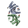
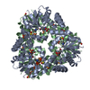
- Assembly
| Deposited unit | 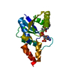
| ||||||||
|---|---|---|---|---|---|---|---|---|---|
| 1 | x 6
| ||||||||
| Unit cell |
| ||||||||
| Components on special symmetry positions |
|
-Components
| #1: Protein | Mass: 17159.738 Da / Num. of mol.: 1 Source method: isolated from a genetically manipulated source Source: (gene. exp.) Mycobacterium tuberculosis (bacteria) / Gene: coaD, kdtB, Rv2965c, MT3043, MTCY349.22, u0002e / Production host: References: UniProt: P0A530, UniProt: P9WPA5*PLUS, pantetheine-phosphate adenylyltransferase |
|---|---|
| #2: Chemical | ChemComp-COA /   Mass: 767.534 Da / Num. of mol.: 1 / Source method: obtained synthetically / Formula: C21H36N7O16P3S Mass: 767.534 Da / Num. of mol.: 1 / Source method: obtained synthetically / Formula: C21H36N7O16P3S |
| #3: Water | ChemComp-HOH /  Mass: 18.015 Da / Num. of mol.: 63 / Source method: isolated from a natural source / Formula: H2O Mass: 18.015 Da / Num. of mol.: 63 / Source method: isolated from a natural source / Formula: H2O |
| Compound details | AUTHORS STATE THAT LIGAND-FERMENT INTERACTION BETWEEN THE STARTING COMPOUND COENZYME A (LIGAND CODE ...AUTHORS STATE THAT LIGAND-FERMENT INTERACTIO |
-Experimental details
-Experiment
| Experiment | Method: X-RAY DIFFRACTION / Number of used crystals: 1 |
|---|
- Sample preparation
| Crystal | Density Matthews: 3.15 Å3/Da / Density % sol: 60.94 % |
|---|
-Data collection
| Diffraction | Mean temperature: 100 K |
|---|---|
| Diffraction source | Source: SYNCHROTRON / Site: SPring-8  / Beamline: BL41XU / Wavelength: 0.8 / Beamline: BL41XU / Wavelength: 0.8 |
| Detector | Type: ADSC QUANTUM 315r / Detector: CCD / Date: Jan 1, 2009 |
| Radiation | Protocol: SINGLE WAVELENGTH / Monochromatic (M) / Laue (L): M / Scattering type: x-ray |
| Radiation wavelength | Wavelength: 0.8 Å / Relative weight: 1 |
| Reflection | Resolution: 1.5→40 Å / Num. obs: 34704 / % possible obs: 98.2 % / Observed criterion σ(I): 2 / Rmerge(I) obs: 0.048 |
- Processing
| Software |
| ||||||||||||||||||||||||
|---|---|---|---|---|---|---|---|---|---|---|---|---|---|---|---|---|---|---|---|---|---|---|---|---|---|
| Refinement | Method to determine structure: MOLECULAR REPLACEMENT / Resolution: 1.59→9.7 Å / Stereochemistry target values: ML
| ||||||||||||||||||||||||
| Refinement step | Cycle: LAST / Resolution: 1.59→9.7 Å
| ||||||||||||||||||||||||
| Refine LS restraints |
|
