 Movie
Movie Controller


+ Open data

- Basic information
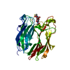
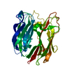
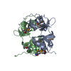
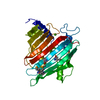
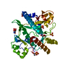
| Entry | Database: PDB / ID: 3pms | ||||||
|---|---|---|---|---|---|---|---|
| Title | Recombinant peptide: N-glycanase F (PNGase F) | ||||||
 Components Components | Peptide:N-glycosidase F | ||||||
 Keywords Keywords | HYDROLASE / Jelly Roll Fold / N-Glycanase | ||||||
| Function / homology |  Function and homology information Function and homology informationoxidoreductase activity, acting on paired donors, with incorporation or reduction of molecular oxygen, reduced ascorbate as one donor, and incorporation of one atom of oxygen / peptide-N4-(N-acetyl-beta-glucosaminyl)asparagine amidase / peptide-N4-(N-acetyl-beta-glucosaminyl)asparagine amidase activity / hydrolase activity, acting on glycosyl bonds Similarity search - Function | ||||||
| Biological species |  Elizabethkingia meningoseptica (bacteria) Elizabethkingia meningoseptica (bacteria) | ||||||
| Method |  X-RAY DIFFRACTION / MOLECULAR REPLACEMENT / molecular replacement / Resolution: 1.57 Å X-RAY DIFFRACTION / MOLECULAR REPLACEMENT / molecular replacement / Resolution: 1.57 Å | ||||||
 Authors Authors | Filitcheva, J. / Anderson, B.F. / Norris, G.E. | ||||||
 Citation Citation | Journal: To be Published Title: Recombinant peptide:N-glycanase F (PNGase F) Authors: Filitcheva, J. / Anderson, B.F. / Norris, G.E. | ||||||
| History |
|
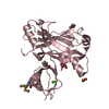
- Structure visualization
| Structure viewer | Molecule: MolmilJmol/JSmol |
|---|
- Downloads & links
-Download
| PDBx/mmCIF format | 3pms.cif.gz | 90 KB | Display | PDBx/mmCIF format |
|---|---|---|---|---|
| PDB format | pdb3pms.ent.gz | 66.3 KB | Display | PDB format |
| PDBx/mmJSON format | 3pms.json.gz | Tree view | PDBx/mmJSON format | |
| Others |  Other downloads Other downloads |
-Validation report
| Arichive directory | https://data.pdbj.org/pub/pdb/validation_reports/pm/3pmsftp://data.pdbj.org/pub/pdb/validation_reports/pm/3pms | HTTPS FTP |
|---|
-Related structure data
| Related structure data | 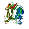 1pgsS S: Starting model for refinement |
|---|---|
| Similar structure data |


-Links
 PDBj
PDBj- Assembly
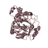
| Deposited unit | 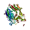
| ||||||||
|---|---|---|---|---|---|---|---|---|---|
| 1 |
| ||||||||
| Unit cell |
|
-Components
| #1: Protein | Mass: 36292.477 Da / Num. of mol.: 1 Source method: isolated from a genetically manipulated source Source: (gene. exp.) Elizabethkingia meningoseptica (bacteria)Strain: CDC 3552 / Gene: png / Plasmid: OPH6 / Production host: References: UniProt: Q9XBM8, peptide-N4-(N-acetyl-beta-glucosaminyl)asparagine amidase | ||||||||
|---|---|---|---|---|---|---|---|---|---|
| #2: Chemical |   Mass: 96.063 Da / Num. of mol.: 2 / Source method: obtained synthetically / Formula: SO4 Mass: 96.063 Da / Num. of mol.: 2 / Source method: obtained synthetically / Formula: SO4#3: Chemical | ChemComp-GOL /   Mass: 92.094 Da / Num. of mol.: 10 / Source method: obtained synthetically / Formula: C3H8O3 Mass: 92.094 Da / Num. of mol.: 10 / Source method: obtained synthetically / Formula: C3H8O3#4: Chemical |   Mass: 59.044 Da / Num. of mol.: 2 / Source method: obtained synthetically / Formula: C2H3O2 Mass: 59.044 Da / Num. of mol.: 2 / Source method: obtained synthetically / Formula: C2H3O2#5: Water | ChemComp-HOH / |  Mass: 18.015 Da / Num. of mol.: 424 / Source method: isolated from a natural source / Formula: H2O Mass: 18.015 Da / Num. of mol.: 424 / Source method: isolated from a natural source / Formula: H2OHas protein modification | Y | |
-Experimental details
-Experiment
| Experiment | Method: X-RAY DIFFRACTION / Number of used crystals: 1 |
|---|
- Sample preparation
| Crystal | Density Matthews: 2.31 Å3/Da / Density % sol: 46.7 % |
|---|---|
| Crystal grow | Temperature: 295.15 K / Method: vapor diffusion, hanging drop / pH: 4.5 Details: 25% PEG 4000, 0.2M ammonium sulfate, 0.1M sodium acetate, pH 4.5, VAPOR DIFFUSION, HANGING DROP, temperature 295.15K |
-Data collection
| Diffraction | Mean temperature: 120 K |
|---|---|
| Diffraction source | Source: ROTATING ANODE / Type: RIGAKU MICROMAX-007 / Wavelength: 1.542 Å |
| Detector | Type: RIGAKU RAXIS IV++ / Detector: IMAGE PLATE / Date: Jul 10, 2009 |
| Radiation | Monochromator: Capillary optics / Protocol: SINGLE WAVELENGTH / Monochromatic (M) / Laue (L): M / Scattering type: x-ray |
| Radiation wavelength | Wavelength: 1.542 Å / Relative weight: 1 |
| Reflection | Resolution: 1.57→40.41 Å / Num. all: 43449 / Num. obs: 41972 / % possible obs: 96.6 % / Rmerge(I) obs: 0.057 |
-Phasing
| Phasing | Method: molecular replacement |
|---|
- Processing
| Software |
| |||||||||||||||||||||||||||||||||||||||||||||||||||||||||||||||||||||||||||||||||||||
|---|---|---|---|---|---|---|---|---|---|---|---|---|---|---|---|---|---|---|---|---|---|---|---|---|---|---|---|---|---|---|---|---|---|---|---|---|---|---|---|---|---|---|---|---|---|---|---|---|---|---|---|---|---|---|---|---|---|---|---|---|---|---|---|---|---|---|---|---|---|---|---|---|---|---|---|---|---|---|---|---|---|---|---|---|---|---|
| Refinement | Method to determine structure: MOLECULAR REPLACEMENT Starting model: PDB ENTRY 1PGS Resolution: 1.57→34.02 Å / Cor.coef. Fo:Fc: 0.97 / Cor.coef. Fo:Fc free: 0.958 / Occupancy max: 1 / Occupancy min: 0.2 / SU B: 1.281 / SU ML: 0.047 / Cross valid method: THROUGHOUT / σ(F): 0 / ESU R Free: 0.076 / Stereochemistry target values: MAXIMUM LIKELIHOOD Details: HYDROGENS HAVE BEEN ADDED IN THE RIDING POSITIONS U VALUES: REFINED INDIVIDUALLY
| |||||||||||||||||||||||||||||||||||||||||||||||||||||||||||||||||||||||||||||||||||||
| Solvent computation | Ion probe radii: 0.8 Å / Shrinkage radii: 0.8 Å / VDW probe radii: 1.4 Å / Solvent model: MASK | |||||||||||||||||||||||||||||||||||||||||||||||||||||||||||||||||||||||||||||||||||||
| Displacement parameters | Biso max: 55.05 Å2 / Biso mean: 18.3064 Å2 / Biso min: 6.03 Å2
| |||||||||||||||||||||||||||||||||||||||||||||||||||||||||||||||||||||||||||||||||||||
| Refinement step | Cycle: LAST / Resolution: 1.57→34.02 Å
| |||||||||||||||||||||||||||||||||||||||||||||||||||||||||||||||||||||||||||||||||||||
| Refine LS restraints |
| |||||||||||||||||||||||||||||||||||||||||||||||||||||||||||||||||||||||||||||||||||||
| LS refinement shell | Resolution: 1.57→1.611 Å / Total num. of bins used: 20
|
