 Movie
Movie Controller

[English] 日本語
 Yorodumi
- PDB-1rpq: High Affinity IgE Receptor (alpha chain) Complexed with Tight-Bin... -
+ Open data

- Basic information
| Entry | Database: PDB / ID: 1rpq | |||||||||
|---|---|---|---|---|---|---|---|---|---|---|
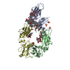
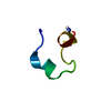
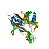
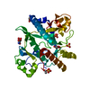
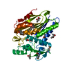
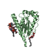
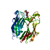
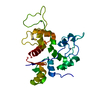
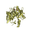
| Title | High Affinity IgE Receptor (alpha chain) Complexed with Tight-Binding E131 'zeta' Peptide from Phage Display | |||||||||
 Components Components |
| |||||||||
 Keywords Keywords | MEMBRANE PROTEIN / receptor-peptide complex | |||||||||
| Function / homology |  Function and homology information Function and homology informationhigh-affinity IgE receptor activity / type I hypersensitivity / eosinophil degranulation / IgE binding / Fc epsilon receptor (FCERI) signaling / type 2 immune response / mast cell degranulation / immunoglobulin mediated immune response / Role of LAT2/NTAL/LAB on calcium mobilization / FCERI mediated Ca+2 mobilization ...high-affinity IgE receptor activity / type I hypersensitivity / eosinophil degranulation / IgE binding / Fc epsilon receptor (FCERI) signaling / type 2 immune response / mast cell degranulation / immunoglobulin mediated immune response / Role of LAT2/NTAL/LAB on calcium mobilization / FCERI mediated Ca+2 mobilization / FCERI mediated MAPK activation / FCERI mediated NF-kB activation / cell surface receptor signaling pathway / external side of plasma membrane / cell surface / plasma membrane Similarity search - Function | |||||||||
| Biological species |  Homo sapiens (human) Homo sapiens (human) | |||||||||
| Method |  X-RAY DIFFRACTION / SYNCHROTRON / MOLECULAR REPLACEMENT / Resolution: 3 Å X-RAY DIFFRACTION / SYNCHROTRON / MOLECULAR REPLACEMENT / Resolution: 3 Å | |||||||||
 Authors Authors | Stamos, J. / Eigenbrot, C. / Nakamura, G.R. / Reynolds, M.E. / Yin, J.P. / Lowman, H.B. / Fairbrother, W.J. / Starovasnik, M.A. | |||||||||
 Citation Citation | Journal: Structure / Year: 2004 Title: Convergent Recognition of the IgE Binding Site on the High-Affinity IgE Receptor. Authors: Stamos, J. / Eigenbrot, C. / Nakamura, G.R. / Reynolds, M.E. / Yin, J.P. / Lowman, H.B. / Fairbrother, W.J. / Starovasnik, M.A. #1: Journal: Proc.Natl.Acad.Sci.USA / Year: 2002Title: Stable "Zeta" Peptides that Act as Potent Antagonists of the High-Affinity Ige Receptor Authors: Nakamura, G.R. / Reynolds, M.E. / Chen, Y.M. / Starovasnik, M.A. / Lowman, H.B. | |||||||||
| History |
|
- Structure visualization
| Structure viewer | Molecule: MolmilJmol/JSmol |
|---|
- Downloads & links
-Download
| PDBx/mmCIF format | 1rpq.cif.gz | 178.7 KB | Display | PDBx/mmCIF format |
|---|---|---|---|---|
| PDB format | pdb1rpq.ent.gz | 146.6 KB | Display | PDB format |
| PDBx/mmJSON format | 1rpq.json.gz | Tree view | PDBx/mmJSON format | |
| Others |  Other downloads Other downloads |
-Validation report
| Arichive directory | https://data.pdbj.org/pub/pdb/validation_reports/rp/1rpqftp://data.pdbj.org/pub/pdb/validation_reports/rp/1rpq | HTTPS FTP |
|---|
-Related structure data
| Related structure data | 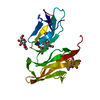 1f2qS S: Starting model for refinement |
|---|---|
| Similar structure data |


-Links
 PDBj
PDBj


- Assembly
| Deposited unit | 
| |||||||||||||||
|---|---|---|---|---|---|---|---|---|---|---|---|---|---|---|---|---|
| 1 | 
| |||||||||||||||
| 2 | 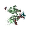
| |||||||||||||||
| 3 | 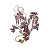
| |||||||||||||||
| 4 | 
| |||||||||||||||
| 5 | 
| |||||||||||||||
| 6 | 
| |||||||||||||||
| Unit cell |
| |||||||||||||||
| Noncrystallographic symmetry (NCS) | NCS domain:
|
-Components
-Protein / Protein/peptide , 2 types, 8 molecules ABCDWXYZ
| #1: Protein | Mass: 20462.738 Da / Num. of mol.: 4 / Fragment: alpha chain extracellular domains Source method: isolated from a genetically manipulated source Source: (gene. exp.) Homo sapiens (human) / Gene: FCER1A, FCE1A / Plasmid: pAcGP67B / Cell line (production host): SF9 / Production host:   Spodoptera frugiperda (fall armyworm) / References: UniProt: P12319 Spodoptera frugiperda (fall armyworm) / References: UniProt: P12319#2: Protein/peptide | Mass: 2539.898 Da / Num. of mol.: 4 / Source method: obtained synthetically Details: Random peptide sequences displayed on phage, selected for binding to Fc(epsilon)RI(alpha) |
|---|
-Sugars , 5 types, 16 molecules 
| #3: Polysaccharide |   Source method: isolated from a genetically manipulated source Details: oligosaccharide / References: triacetyl-beta-chitotriose #4: Polysaccharide | beta-D-mannopyranose-(1-3)-[beta-D-mannopyranose-(1-4)]beta-D-mannopyranose-(1-4)-2-acetamido-2- ...beta-D-mannopyranose-(1-3)-[beta-D-mannopyranose-(1-4)]beta-D-mannopyranose-(1-4)-2-acetamido-2-deoxy-beta-D-glucopyranose-(1-4)-2-acetamido-2-deoxy-beta-D-glucopyranose Source method: isolated from a genetically manipulated source #5: Polysaccharide | beta-D-mannopyranose-(1-4)-2-acetamido-2-deoxy-beta-D-glucopyranose-(1-4)-2-acetamido-2-deoxy-beta- ...beta-D-mannopyranose-(1-4)-2-acetamido-2-deoxy-beta-D-glucopyranose-(1-4)-2-acetamido-2-deoxy-beta-D-glucopyranose Source method: isolated from a genetically manipulated source #6: Polysaccharide | Source method: isolated from a genetically manipulated source #7: Sugar | ChemComp-NAG /  Type: D-saccharide, beta linking / Mass: 221.208 Da / Num. of mol.: 4 Type: D-saccharide, beta linking / Mass: 221.208 Da / Num. of mol.: 4Source method: isolated from a genetically manipulated source Formula: C8H15NO6 |
|---|
-Non-polymers , 2 types, 4 molecules 

| #8: Chemical |  Mass: 96.063 Da / Num. of mol.: 2 / Source method: obtained synthetically / Formula: SO4 Mass: 96.063 Da / Num. of mol.: 2 / Source method: obtained synthetically / Formula: SO4#9: Chemical |  Mass: 192.124 Da / Num. of mol.: 2 / Source method: obtained synthetically / Formula: C6H8O7 Mass: 192.124 Da / Num. of mol.: 2 / Source method: obtained synthetically / Formula: C6H8O7 |
|---|
-Details
| Has protein modification | Y |
|---|
-Experimental details
-Experiment
| Experiment | Method: X-RAY DIFFRACTION / Number of used crystals: 1 |
|---|
- Sample preparation
| Crystal | Density Matthews: 4.23 Å3/Da / Density % sol: 70.89 % |
|---|---|
| Crystal grow | Temperature: 292 K / Method: vapor diffusion, hanging drop / pH: 4.2 Details: PEG 2000 monomethyl ether, ammonium sulfate, sodium citrate, pH 4.2, VAPOR DIFFUSION, HANGING DROP, temperature 292K |
-Data collection
| Diffraction | Mean temperature: 100 K |
|---|---|
| Diffraction source | Source: SYNCHROTRON / Site: SSRL  / Beamline: BL9-2 / Wavelength: 1.033 Å / Beamline: BL9-2 / Wavelength: 1.033 Å |
| Detector | Type: ADSC QUANTUM 4 / Detector: CCD / Date: Jan 5, 2001 |
| Radiation | Monochromator: double crystal / Protocol: SINGLE WAVELENGTH / Monochromatic (M) / Laue (L): M / Scattering type: x-ray |
| Radiation wavelength | Wavelength: 1.033 Å / Relative weight: 1 |
| Reflection | Resolution: 3→30 Å / Num. all: 30473 / Num. obs: 30473 / % possible obs: 96.5 % / Observed criterion σ(F): -10 / Observed criterion σ(I): -3 / Redundancy: 4.9 % / Biso Wilson estimate: 85.3 Å2 / Rmerge(I) obs: 0.068 / Net I/σ(I): 7.4 |
| Reflection shell | Resolution: 3→3.11 Å / Redundancy: 4.7 % / Rmerge(I) obs: 0.69 / Mean I/σ(I) obs: 2 / Num. unique all: 2644 / % possible all: 85 |
- Processing
| Software |
| |||||||||||||||||||||||||||||||||||||||||||||||||||||||||||||||
|---|---|---|---|---|---|---|---|---|---|---|---|---|---|---|---|---|---|---|---|---|---|---|---|---|---|---|---|---|---|---|---|---|---|---|---|---|---|---|---|---|---|---|---|---|---|---|---|---|---|---|---|---|---|---|---|---|---|---|---|---|---|---|---|---|
| Refinement | Method to determine structure: MOLECULAR REPLACEMENT Starting model: PDB entry 1F2Q Resolution: 3→30 Å / Rfactor Rfree error: 0.011 / Data cutoff high absF: 1000000 / Data cutoff low absF: 0.001 / Isotropic thermal model: RESTRAINED / Cross valid method: THROUGHOUT / σ(F): 0.2 / Stereochemistry target values: Engh & Huber / Details: BULK SOLVENT MODEL USED
| |||||||||||||||||||||||||||||||||||||||||||||||||||||||||||||||
| Displacement parameters | Biso mean: 70.4 Å2
| |||||||||||||||||||||||||||||||||||||||||||||||||||||||||||||||
| Refine analyze |
| |||||||||||||||||||||||||||||||||||||||||||||||||||||||||||||||
| Refinement step | Cycle: LAST / Resolution: 3→30 Å
| |||||||||||||||||||||||||||||||||||||||||||||||||||||||||||||||
| Refine LS restraints |
| |||||||||||||||||||||||||||||||||||||||||||||||||||||||||||||||
| Refine LS restraints NCS | Refine-ID: X-RAY DIFFRACTION / Rms dev Biso : 2.7 Å2 / Weight Biso : 3 / Weight position: 1000
| |||||||||||||||||||||||||||||||||||||||||||||||||||||||||||||||
| LS refinement shell | Refine-ID: X-RAY DIFFRACTION / Total num. of bins used: 10
| |||||||||||||||||||||||||||||||||||||||||||||||||||||||||||||||
| Xplor file |
|
