translation elongation factor activity / regulation of DNA-templated transcription elongation / transcription elongation by RNA polymerase II / structural constituent of ribosome / ribosome / mRNA binding / regulation of transcription by RNA polymerase II / regulation of DNA-templated transcription / zinc ion binding Similarity search - Function
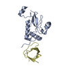
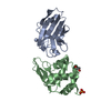
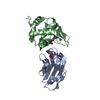
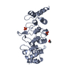
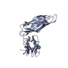
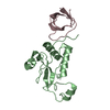
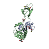
Journal: Proc Natl Acad Sci U S A / Year: 2011 Title: RNA polymerase and transcription elongation factor Spt4/5 complex structure. Authors: Brianna J Klein / Daniel Bose / Kevin J Baker / Zahirah M Yusoff / Xiaodong Zhang / Katsuhiko S Murakami / Abstract: Spt4/5 in archaea and eukaryote and its bacterial homolog NusG is the only elongation factor conserved in all three domains of life and plays many key roles in cotranscriptional regulation and in ...Spt4/5 in archaea and eukaryote and its bacterial homolog NusG is the only elongation factor conserved in all three domains of life and plays many key roles in cotranscriptional regulation and in recruiting other factors to the elongating RNA polymerase. Here, we present the crystal structure of Spt4/5 as well as the structure of RNA polymerase-Spt4/5 complex using cryoelectron microscopy reconstruction and single particle analysis. The Spt4/5 binds in the middle of RNA polymerase claw and encloses the DNA, reminiscent of the DNA polymerase clamp and ring helicases. The transcription elongation complex model reveals that the Spt4/5 is an upstream DNA holder and contacts the nontemplate DNA in the transcription bubble. These structures reveal that the cellular RNA polymerases also use a strategy of encircling DNA to enhance its processivity as commonly observed for many nucleic acid processing enzymes including DNA polymerases and helicases.
Mass: 18.015 Da / Num. of mol.: 111 / Source method: isolated from a natural source / Formula: H2O
-
Experimental details
-
Experiment
Experiment
Method: X-RAY DIFFRACTION / Number of used crystals: 1
-
Sample preparation
Crystal
Density Matthews: 2.27 Å3/Da / Density % sol: 45.72 %
Crystal grow
Temperature: 295 K / Method: vapor diffusion, sitting drop / pH: 5.5 Details: 0.1 M Bis-Tris (pH 5.5), 0.2 M NaCl, and 25% PEG3350, VAPOR DIFFUSION, SITTING DROP, temperature 295K
-
Data collection
Diffraction source
Source: SYNCHROTRON / Site: CHESS / Beamline: F1 / Wavelength: 0.9181 Å
Protocol: SINGLE WAVELENGTH / Monochromatic (M) / Laue (L): M / Scattering type: x-ray
Radiation wavelength
Wavelength: 0.9181 Å / Relative weight: 1
Reflection
Resolution: 1.8→30 Å / Num. obs: 44007
-
Processing
Software
Name
Version
Classification
PHASER
phasing
REFMAC
5.5.0072
refinement
HKL-2000
datareduction
HKL-2000
datascaling
Refinement
Method to determine structure: MOLECULAR REPLACEMENT / Resolution: 1.8→30 Å / Cor.coef. Fo:Fc: 0.945 / Cor.coef. Fo:Fc free: 0.911 / SU B: 4.424 / SU ML: 0.132 / Cross valid method: THROUGHOUT / ESU R Free: 0.154 / Stereochemistry target values: MAXIMUM LIKELIHOOD / Details: HYDROGENS HAVE BEEN ADDED IN THE RIDING POSITIONS
Rfactor
Num. reflection
% reflection
Selection details
Rfree
0.29234
2208
5 %
RANDOM
Rwork
0.23644
-
-
-
obs
0.23928
41656
98.13 %
-
Solvent computation
Ion probe radii: 0.8 Å / Shrinkage radii: 0.8 Å / VDW probe radii: 1.4 Å / Solvent model: MASK
Displacement parameters
Biso mean: 31.098 Å2
Baniso -1
Baniso -2
Baniso -3
1-
0.46 Å2
0 Å2
0 Å2
2-
-
1.09 Å2
0 Å2
3-
-
-
-1.55 Å2
Refinement step
Cycle: LAST / Resolution: 1.8→30 Å
Protein
Nucleic acid
Ligand
Solvent
Total
Num. atoms
3238
0
56
111
3405
Refine LS restraints
Refine-ID
Type
Dev ideal
Dev ideal target
Number
X-RAY DIFFRACTION
r_bond_refined_d
0.024
0.022
3343
X-RAY DIFFRACTION
r_bond_other_d
X-RAY DIFFRACTION
r_angle_refined_deg
1.991
1.988
4499
X-RAY DIFFRACTION
r_angle_other_deg
X-RAY DIFFRACTION
r_dihedral_angle_1_deg
6.246
5
410
X-RAY DIFFRACTION
r_dihedral_angle_2_deg
31.936
23.433
134
X-RAY DIFFRACTION
r_dihedral_angle_3_deg
18.641
15
598
X-RAY DIFFRACTION
r_dihedral_angle_4_deg
20.64
15
24
X-RAY DIFFRACTION
r_chiral_restr
0.14
0.2
512
X-RAY DIFFRACTION
r_gen_planes_refined
0.01
0.021
2434
X-RAY DIFFRACTION
r_gen_planes_other
X-RAY DIFFRACTION
r_nbd_refined
X-RAY DIFFRACTION
r_nbd_other
X-RAY DIFFRACTION
r_nbtor_refined
X-RAY DIFFRACTION
r_nbtor_other
X-RAY DIFFRACTION
r_xyhbond_nbd_refined
X-RAY DIFFRACTION
r_xyhbond_nbd_other
X-RAY DIFFRACTION
r_metal_ion_refined
X-RAY DIFFRACTION
r_metal_ion_other
X-RAY DIFFRACTION
r_symmetry_vdw_refined
X-RAY DIFFRACTION
r_symmetry_vdw_other
X-RAY DIFFRACTION
r_symmetry_hbond_refined
X-RAY DIFFRACTION
r_symmetry_hbond_other
X-RAY DIFFRACTION
r_symmetry_metal_ion_refined
X-RAY DIFFRACTION
r_symmetry_metal_ion_other
X-RAY DIFFRACTION
r_mcbond_it
1.311
1.5
2062
X-RAY DIFFRACTION
r_mcbond_other
X-RAY DIFFRACTION
r_mcangle_it
2.244
2
3360
X-RAY DIFFRACTION
r_scbond_it
3.6
3
1281
X-RAY DIFFRACTION
r_scangle_it
5.983
4.5
1139
X-RAY DIFFRACTION
r_rigid_bond_restr
X-RAY DIFFRACTION
r_sphericity_free
X-RAY DIFFRACTION
r_sphericity_bonded
LS refinement shell
Resolution: 1.8→1.847 Å / Total num. of bins used: 20
Rfactor
Num. reflection
% reflection
Rfree
0.407
142
-
Rwork
0.396
2684
-
obs
-
-
86.26 %
+
About Yorodumi
-
News
-
Feb 9, 2022. New format data for meta-information of EMDB entries
New format data for meta-information of EMDB entries
Version 3 of the EMDB header file is now the official format.
The previous official version 1.9 will be removed from the archive.
In the structure databanks used in Yorodumi, some data are registered as the other names, "COVID-19 virus" and "2019-nCoV". Here are the details of the virus and the list of structure data.
Jan 31, 2019. EMDB accession codes are about to change! (news from PDBe EMDB page)
EMDB accession codes are about to change! (news from PDBe EMDB page)
The allocation of 4 digits for EMDB accession codes will soon come to an end. Whilst these codes will remain in use, new EMDB accession codes will include an additional digit and will expand incrementally as the available range of codes is exhausted. The current 4-digit format prefixed with “EMD-” (i.e. EMD-XXXX) will advance to a 5-digit format (i.e. EMD-XXXXX), and so on. It is currently estimated that the 4-digit codes will be depleted around Spring 2019, at which point the 5-digit format will come into force.
The EM Navigator/Yorodumi systems omit the EMD- prefix.
Related info.:Q: What is EMD? / ID/Accession-code notation in Yorodumi/EM Navigator
Yorodumi is a browser for structure data from EMDB, PDB, SASBDB, etc.
This page is also the successor to EM Navigator detail page, and also detail information page/front-end page for Omokage search.
The word "yorodu" (or yorozu) is an old Japanese word meaning "ten thousand". "mi" (miru) is to see.
Related info.:EMDB / PDB / SASBDB / Comparison of 3 databanks / Yorodumi Search / Aug 31, 2016. New EM Navigator & Yorodumi / Yorodumi Papers / Jmol/JSmol / Function and homology information / Changes in new EM Navigator and Yorodumi
 Movie
Movie Controller

 Yorodumi
 Open data
 Basic information
 Components
 Keywords
 Function and homology information
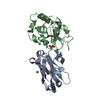
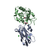
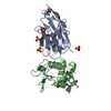
 Pyrococcus furiosus (archaea)
 X-RAY DIFFRACTION /
  Authors
 Citation
 Structure visualization
 Downloads & links
 Other downloads
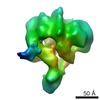

 PDBj
PDBj


 Assembly
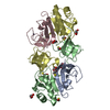


 Mass: 65.409 Da / Num. of mol.: 2 / Source method: obtained synthetically / Formula: Zn
 Mass: 78.133 Da / Num. of mol.: 6 / Source method: obtained synthetically / Formula: C2H6OS
 Mass: 92.094 Da / Num. of mol.: 5 / Source method: obtained synthetically / Formula: C3H8O3
 Sample preparation
 Processing