 Movie
Movie Controller

[English] 日本語
 Yorodumi
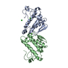
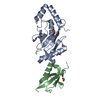
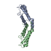
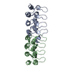
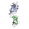
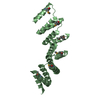
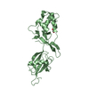
- PDB-3p2p: ENHANCED ACTIVITY AND ALTERED SPECIFICITY OF PHOSPHOLIPASE A2 BY ... -
+ Open data

- Basic information
| Entry | Database: PDB / ID: 3p2p | ||||||
|---|---|---|---|---|---|---|---|
| Title | ENHANCED ACTIVITY AND ALTERED SPECIFICITY OF PHOSPHOLIPASE A2 BY DELETION OF A SURFACE LOOP | ||||||
 Components Components | PHOSPHOLIPASE A2 | ||||||
 Keywords Keywords | HYDROLASE(CARBOXYL ESTER) | ||||||
| Function / homology |  Function and homology information Function and homology informationregulation of D-glucose import across plasma membrane / positive regulation of podocyte apoptotic process / phosphatidylglycerol metabolic process / phosphatidylcholine metabolic process / A2-type glycerophospholipase activity / leukotriene biosynthetic process / : / bile acid binding / phospholipase A2 / neutrophil mediated immunity ...regulation of D-glucose import across plasma membrane / positive regulation of podocyte apoptotic process / phosphatidylglycerol metabolic process / phosphatidylcholine metabolic process / A2-type glycerophospholipase activity / leukotriene biosynthetic process / : / bile acid binding / phospholipase A2 / neutrophil mediated immunity / positive regulation of calcium ion transport into cytosol / lipid catabolic process / neutrophil chemotaxis / positive regulation of interleukin-8 production / phospholipid binding / positive regulation of immune response / positive regulation of fibroblast proliferation / fatty acid biosynthetic process / cellular response to insulin stimulus / positive regulation of MAPK cascade / intracellular signal transduction / signaling receptor binding / calcium ion binding / positive regulation of cell population proliferation / cell surface / positive regulation of transcription by RNA polymerase II / extracellular region Similarity search - Function | ||||||
| Biological species |  | ||||||
| Method |  X-RAY DIFFRACTION / Resolution: 2.1 Å X-RAY DIFFRACTION / Resolution: 2.1 Å | ||||||
 Authors Authors | Dijkstra, B.W. / Thunnissen, M.M.G.M. / Kalk, K.H. / Drenth, J. | ||||||
 Citation Citation | Journal: Science / Year: 1989 Title: Enhanced activity and altered specificity of phospholipase A2 by deletion of a surface loop. Authors: Kuipers, O.P. / Thunnissen, M.M. / de Geus, P. / Dijkstra, B.W. / Drenth, J. / Verheij, H.M. / de Haas, G.H. #1: Journal: J.Mol.Biol. / Year: 1983Title: Structure of Porcine Pancreatic Phospholipase A2 at 2.6 Angstroms Resolution and Comparison with Bovine Phospholipase A2 Authors: Dijkstra, B.W. / Renetseder, R. / Kalk, K.H. / Hol, W.G.J. / Drenth, J. #2: Journal: Acta Crystallogr.,Sect.B / Year: 1982Title: The Structure of Bovine Pancreatic Prophospholipase A2 at 3.0 Angstroms Resolution Authors: Dijkstra, B.W. / Vannes, G.J.H. / Kalk, K.H. / Brandenburg, N.P. / Hol, W.G.J. / Drenth, J. #3: Journal: J.Mol.Biol. / Year: 1981Title: Structure of Bovine Pancreatic Phospholipase A2 at 1.7 Angstroms Resolution Authors: Dijkstra, B.W. / Kalk, K.H. / Hol, W.G.J. / Drenth, J. #4: Journal: Nature / Year: 1981Title: Active Site and Catalytic Mechanism of Phospholipase A2 Authors: Dijkstra, B.W. / Drenth, J. / Kalk, K.H. #5: Journal: J.Mol.Biol. / Year: 1978Title: Three-Dimensional Structure and Disulfide Bond Connections in Bovine Pancreatic Phospholipase A2 Authors: Dijkstra, B.W. / Drenth, J. / Kalk, K.H. / Vandermaelen, P.J. | ||||||
| History |
|
- Structure visualization
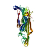
| Structure viewer | Molecule: MolmilJmol/JSmol |
|---|
- Downloads & links
-Download
| PDBx/mmCIF format | 3p2p.cif.gz | 64.2 KB | Display | PDBx/mmCIF format |
|---|---|---|---|---|
| PDB format | pdb3p2p.ent.gz | 46.8 KB | Display | PDB format |
| PDBx/mmJSON format | 3p2p.json.gz | Tree view | PDBx/mmJSON format | |
| Others |  Other downloads Other downloads |
-Validation report
| Arichive directory | https://data.pdbj.org/pub/pdb/validation_reports/p2/3p2pftp://data.pdbj.org/pub/pdb/validation_reports/p2/3p2p | HTTPS FTP |
|---|
-Related structure data
| Similar structure data |
|---|

-Links
 PDBj
PDBj

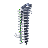
- Assembly
| Deposited unit | 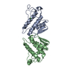
| ||||||||
|---|---|---|---|---|---|---|---|---|---|
| 1 |
| ||||||||
| Unit cell |
| ||||||||
| Atom site foot note | 1: SEE REMARK 6. | ||||||||
| Noncrystallographic symmetry (NCS) | NCS oper: (Code: given Matrix: (-0.311854, -0.931273, -0.188355), Vector: Details | THE ENZYME CRYSTALLIZES WITH A DIMER IN THE ASYMMETRIC UNIT. THE TRANSFORMATION PRESENTED ON *MTRIX 1* RECORDS BELOW CAN BE APPLIED TO SUBUNIT 2 (CHAIN *B*) TO OBTAIN OPTIMAL SUPERPOSITION OF C-ALPHA COORDINATES ON THOSE OF SUBUNIT 1 (CHAIN *A*). THE RMS DIFFERENCE FOR ALL 119 CARBON-ALPHA PAIRS IS 0.542 ANGSTROMS. | |
-Components
| #1: Protein | Mass: 13397.018 Da / Num. of mol.: 2 Source method: isolated from a genetically manipulated source Source: (gene. exp.) #2: Chemical |   Mass: 40.078 Da / Num. of mol.: 3 / Source method: obtained synthetically / Formula: Ca Mass: 40.078 Da / Num. of mol.: 3 / Source method: obtained synthetically / Formula: Ca#3: Water | ChemComp-HOH / |  Mass: 18.015 Da / Num. of mol.: 205 / Source method: isolated from a natural source / Formula: H2O Mass: 18.015 Da / Num. of mol.: 205 / Source method: isolated from a natural source / Formula: H2OHas protein modification | Y | Nonpolymer details | CALCIUM CA B 2 IS BOUND BETWEEN THE *A* AND *B* CHAINS. | |
|---|
-Experimental details
-Experiment
| Experiment | Method: X-RAY DIFFRACTION |
|---|
- Sample preparation
| Crystal | Density Matthews: 2.23 Å3/Da / Density % sol: 44.74 % | ||||||||||||||||||||||||
|---|---|---|---|---|---|---|---|---|---|---|---|---|---|---|---|---|---|---|---|---|---|---|---|---|---|
| Crystal grow | *PLUS pH: 7 / Method: vapor diffusion, hanging drop | ||||||||||||||||||||||||
| Components of the solutions | *PLUS
|
-Data collection
| Reflection | *PLUS Highest resolution: 2.5 Å / % possible obs: 88.5 % |
|---|
- Processing
| Software | Name: TNT / Classification: refinement | ||||||||||||
|---|---|---|---|---|---|---|---|---|---|---|---|---|---|
| Refinement | Rfactor obs: 0.186 / Highest resolution: 2.1 Å | ||||||||||||
| Refine analyze | Luzzati coordinate error obs: 0.25 Å | ||||||||||||
| Refinement step | Cycle: LAST / Highest resolution: 2.1 Å
| ||||||||||||
| Refinement | *PLUS Highest resolution: 2.5 Å / Rfactor obs: 0.237 / Rfactor Rwork: 0.237 | ||||||||||||
| Solvent computation | *PLUS | ||||||||||||
| Displacement parameters | *PLUS | ||||||||||||
| Refine LS restraints | *PLUS Type: bond_d / Dev ideal: 0.009 |
