[English] 日本語
Yorodumi
- PDB-1ymp: The Crystal Structure of a Partial Mouse Notch-1 Ankyrin Domain: ... -

+
Open data


ID or keywords:

Loading...

-
Basic information

Entry
Database: PDB / ID: 1ymp
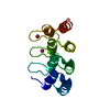
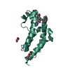
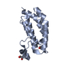
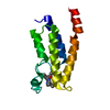
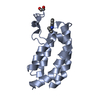
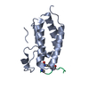
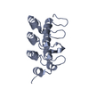
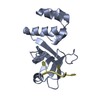
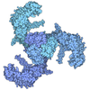
TitleThe Crystal Structure of a Partial Mouse Notch-1 Ankyrin Domain: Repeats 4 Through 7 Preserve an Ankyrin Fold
ComponentsNotch 1 protein
KeywordsTRANSCRIPTION / ankyrin repeats / helix-turn-helix
Function / homology
Function and homology information


Notch signaling involved in heart development / Pre-NOTCH Processing in Golgi / cardiac right ventricle formation / regulation of inner ear auditory receptor cell differentiation / osteoblast fate commitment / negative regulation of inner ear auditory receptor cell differentiation / positive regulation of aorta morphogenesis / negative regulation of photoreceptor cell differentiation / apoptotic process involved in embryonic digit morphogenesis / venous blood vessel morphogenesis ...Notch signaling involved in heart development / Pre-NOTCH Processing in Golgi / cardiac right ventricle formation / regulation of inner ear auditory receptor cell differentiation / osteoblast fate commitment / negative regulation of inner ear auditory receptor cell differentiation / positive regulation of aorta morphogenesis / negative regulation of photoreceptor cell differentiation / apoptotic process involved in embryonic digit morphogenesis / venous blood vessel morphogenesis / regulation of somitogenesis / foregut morphogenesis / regulation of epithelial cell proliferation involved in prostate gland development / positive regulation of ephrin receptor signaling pathway / positive regulation of glial cell differentiation / auditory receptor cell fate commitment / secretory columnal luminar epithelial cell differentiation involved in prostate glandular acinus development / CSL-Notch-Mastermind transcription factor complex / compartment pattern specification / endocardial cushion development / somatic stem cell division / coronary sinus valve morphogenesis / cardiac right atrium morphogenesis / growth involved in heart morphogenesis / regulation of cardioblast proliferation / mesenchymal cell development / cell differentiation in spinal cord / venous endothelial cell differentiation / arterial endothelial cell differentiation / collecting duct development / cell migration involved in endocardial cushion formation / negative regulation of pro-B cell differentiation / prostate gland epithelium morphogenesis / mitral valve formation / : / endocardium morphogenesis / distal tubule development / MAML1-RBP-Jkappa- ICN1 complex / cardiac chamber formation / cardiac atrium morphogenesis / endoderm development / NOTCH1 Intracellular Domain Regulates Transcription / RUNX3 regulates NOTCH signaling / pericardium morphogenesis / atrioventricular node development / cardiac ventricle morphogenesis / positive regulation of transcription of Notch receptor target / negative regulation of endothelial cell chemotaxis / cardiac septum morphogenesis / glomerular mesangial cell development / cellular response to tumor cell / Notch-HLH transcription pathway / positive regulation of smooth muscle cell differentiation / Activated NOTCH1 Transmits Signal to the Nucleus / vasculogenesis involved in coronary vascular morphogenesis / negative regulation of extracellular matrix constituent secretion / regulation of extracellular matrix assembly / chemical synaptic transmission, postsynaptic / positive regulation of apoptotic process involved in morphogenesis / endocardial cell differentiation / left/right axis specification / epithelial to mesenchymal transition involved in endocardial cushion formation / positive regulation of endothelial cell differentiation / cardiac left ventricle morphogenesis / embryonic hindlimb morphogenesis / negative regulation of myotube differentiation / coronary vein morphogenesis / negative regulation of glial cell proliferation / cardiac vascular smooth muscle cell development / neuronal stem cell population maintenance / endocardium development / positive regulation of astrocyte differentiation / cardiac muscle cell myoblast differentiation / negative regulation of cell adhesion molecule production / negative regulation of stem cell differentiation / glial cell differentiation / tissue regeneration / T-helper 17 type immune response / negative regulation of calcium ion-dependent exocytosis / skeletal muscle cell differentiation / positive regulation of cardiac epithelial to mesenchymal transition / regulation of Notch signaling pathway / cardiac epithelial to mesenchymal transition / heart trabecula morphogenesis / embryonic limb morphogenesis / neuron fate commitment / negative regulation of oligodendrocyte differentiation / regulation of cell adhesion involved in heart morphogenesis / interleukin-17-mediated signaling pathway / negative regulation of catalytic activity / negative regulation of myoblast differentiation / camera-type eye morphogenesis / cellular response to follicle-stimulating hormone stimulus / neural tube development / negative regulation of collagen biosynthetic process / negative regulation of cardiac muscle hypertrophy / luteolysis / determination of left/right symmetry / pulmonary valve morphogenesis / tube formation
Similarity search - Function
: / Neurogenic locus notch homolog protein 1 / Notch, C-terminal / Domain of unknown function / : / Notch / Notch, NOD domain / Notch, NODP domain / NOTCH protein / NOTCH protein ...: / Neurogenic locus notch homolog protein 1 / Notch, C-terminal / Domain of unknown function / : / Notch / Notch, NOD domain / Notch, NODP domain / NOTCH protein / NOTCH protein / NOD / NODP / Notch-like domain superfamily / LNR (Lin-12/Notch) repeat profile. / LNR domain / Notch domain / Domain found in Notch and Lin-12 / EGF-like, conserved site / Human growth factor-like EGF / Ankyrin repeat-containing domain / : / Calcium-binding EGF domain / EGF-like domain / EGF-type aspartate/asparagine hydroxylation site / EGF-like calcium-binding, conserved site / Calcium-binding EGF-like domain signature. / Aspartic acid and asparagine hydroxylation site. / EGF-like calcium-binding domain / Calcium-binding EGF-like domain / Epidermal growth factor-like domain. / Ankyrin repeat / EGF-like domain profile. / Growth factor receptor cysteine-rich domain superfamily / EGF-like domain signature 1. / EGF-like domain signature 2. / EGF-like domain / Ankyrin repeat profile. / Ankyrin repeats (3 copies) / Ankyrin repeat region circular profile. / ankyrin repeats / Ankyrin repeat / Ankyrin repeat-containing domain superfamily / Serine Threonine Protein Phosphatase 5, Tetratricopeptide repeat / Alpha Horseshoe / Mainly Alpha
Similarity search - Domain/homology
Neurogenic locus notch homolog protein 1 / Neurogenic locus notch homolog protein 1
Similarity search - Component
Biological speciesMus musculus (house mouse)
MethodX-RAY DIFFRACTION / SYNCHROTRON / MOLECULAR REPLACEMENT / Resolution: 2.2 Å
AuthorsLubman, O.Y. / Kopan, R. / Waksman, G. / Korolev, S.
CitationJournal: Protein Sci. / Year: 2005
Title: The crystal structure of a partial mouse Notch-1 ankyrin domain: repeats 4 through 7 preserve an ankyrin fold.
Authors: Lubman, O.Y. / Kopan, R. / Waksman, G. / Korolev, S.
History
DepositionJan 21, 2005Deposition site: RCSB / Processing site: RCSB
Revision 1.0May 10, 2005Provider: repository / Type: Initial release
Revision 1.1Apr 30, 2008Group: Version format compliance
Revision 1.2Jul 13, 2011Group: Version format compliance
Revision 1.3Aug 23, 2023Group: Data collection / Database references / Refinement description
Category: chem_comp_atom / chem_comp_bond ...chem_comp_atom / chem_comp_bond / database_2 / pdbx_initial_refinement_model
Item: _database_2.pdbx_DOI / _database_2.pdbx_database_accession

-
Structure visualization

Structure viewerMolecule:
MolmilJmol/JSmol

Downloads & links

-
Assembly

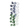
Deposited unit
A: Notch 1 protein
B: Notch 1 protein


Theoretical massNumber of molelcules
Total (without water)30,1562
Polymers30,1562
Non-polymers00
Water2,684149
1
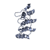
A: Notch 1 protein


Theoretical massNumber of molelcules
Total (without water)15,0781
Polymers15,0781
Non-polymers00
Water181
TypeNameSymmetry operationNumber
identity operation1_555x,y,z1
2
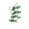
B: Notch 1 protein


Theoretical massNumber of molelcules
Total (without water)15,0781
Polymers15,0781
Non-polymers00
Water181
TypeNameSymmetry operationNumber
identity operation1_555x,y,z1
Unit cell
Length a, b, c (Å)75.789, 75.789, 45.850
Angle α, β, γ (deg.)90.00, 90.00, 120.00
Int Tables number144
Space group name H-MP31
Detailsbiological assembly is a monomer

-
Components

#1: Protein Notch 1 protein


Mass: 15077.961 Da / Num. of mol.: 2 / Fragment: ankryin repeats 3.5-7 (residues 1971-2105)
Source method: isolated from a genetically manipulated source
Source: (gene. exp.) Mus musculus (house mouse) / Gene: Notch1 / Plasmid: pProEX HTc / Production host: Escherichia coli (E. coli) / Strain (production host): JM109 / References: UniProt: Q07008, UniProt: Q01705*PLUS

#2: Water ChemComp-HOH / water


Mass: 18.015 Da / Num. of mol.: 149 / Source method: isolated from a natural source / Formula: H2O

-
Experimental details

-
Experiment

ExperimentMethod: X-RAY DIFFRACTION / Number of used crystals: 1

-
Sample preparation

CrystalDensity Matthews: 2.2 Å3/Da / Density % sol: 48 %
Crystal growTemperature: 298 K / Method: vapor diffusion, hanging drop / pH: 6.5
Details: PEG 10000, cacodylate, pH 6.5, VAPOR DIFFUSION, HANGING DROP, temperature 298K

-
Data collection

DiffractionMean temperature: 98 K
Diffraction sourceSource: SYNCHROTRON / Site: APS / Beamline: 19-BM / Wavelength: 0.9 Å
RadiationProtocol: SINGLE WAVELENGTH / Monochromatic (M) / Laue (L): M / Scattering type: x-ray
Radiation wavelengthWavelength: 0.9 Å / Relative weight: 1
ReflectionResolution: 2.2→50 Å / Num. obs: 14979 / % possible obs: 85.6 % / Observed criterion σ(F): 1 / Observed criterion σ(I): 1 / Rmerge(I) obs: 0.043 / Rsym value: 0.047
Reflection shellResolution: 2.2→2.34 Å / % possible all: 85.6

-
Phasing

Phasing MRCor.coef. Fo:Fc: 0.516 / Packing: 0.515
Highest resolutionLowest resolutionMethodReflection percentσ(F)
Rotation3.5 Å15 Åfast direct97.4 0
Translation4 Å15 Ågeneral: ! target for PC-refinement= e2e298.7 0

-
Processing

Software
NameVersionClassificationNB
CNSrefinement
HKL-2000data reduction
SCALEPACKdata scaling
CNS1.1phasing
RefinementMethod to determine structure: MOLECULAR REPLACEMENT
Starting model: pdb entry 1N0R
Resolution: 2.2→21.88 Å / Data cutoff high absF: 10000 / Data cutoff low absF: 0 / σ(F): 0 / Stereochemistry target values: Engh & Huber
Details: refined as perfect merohedral twin using CNS scripts
RfactorNum. reflection% reflectionSelection details
Rfree0.309 549 3.7 %random
Rwork0.206 ---
obs-11012 73.6 %-
Displacement parametersBiso mean: 73.912 Å2
Baniso -1Baniso -2Baniso -3
1--0.323 Å23.078 Å20 Å2
2---0.323 Å20 Å2
3---0.645 Å2
Refinement stepCycle: LAST / Resolution: 2.2→21.88 Å
ProteinNucleic acidLigandSolventTotal
Num. atoms1949 0 0 149 2098
Refine LS restraints
Refine-IDTypeDev ideal
X-RAY DIFFRACTIONx_bond_d0.011
X-RAY DIFFRACTIONx_angle_deg1.69

+
About Yorodumi

-
News

-
Feb 9, 2022. New format data for meta-information of EMDB entries

New format data for meta-information of EMDB entries

  • Version 3 of the EMDB header file is now the official format.
  • The previous official version 1.9 will be removed from the archive.

Related info.:EMDB header

External links:wwPDB to switch to version 3 of the EMDB data model

-
Aug 12, 2020. Covid-19 info

Covid-19 info

URL: https://pdbj.org/emnavi/covid19.php

New page: Covid-19 featured information page in EM Navigator.

Related info.:Covid-19 info / Mar 5, 2020. Novel coronavirus structure data

+
Mar 5, 2020. Novel coronavirus structure data

Novel coronavirus structure data

Related info.:Yorodumi Speices / Aug 12, 2020. Covid-19 info

External links:COVID-19 featured content - PDBj / Molecule of the Month (242):Coronavirus Proteases

+
Jan 31, 2019. EMDB accession codes are about to change! (news from PDBe EMDB page)

EMDB accession codes are about to change! (news from PDBe EMDB page)

  • The allocation of 4 digits for EMDB accession codes will soon come to an end. Whilst these codes will remain in use, new EMDB accession codes will include an additional digit and will expand incrementally as the available range of codes is exhausted. The current 4-digit format prefixed with “EMD-” (i.e. EMD-XXXX) will advance to a 5-digit format (i.e. EMD-XXXXX), and so on. It is currently estimated that the 4-digit codes will be depleted around Spring 2019, at which point the 5-digit format will come into force.
  • The EM Navigator/Yorodumi systems omit the EMD- prefix.

Related info.:Q: What is EMD? / ID/Accession-code notation in Yorodumi/EM Navigator

External links:EMDB Accession Codes are Changing Soon! / Contact to PDBj

+
Jul 12, 2017. Major update of PDB

Major update of PDB

  • wwPDB released updated PDB data conforming to the new PDBx/mmCIF dictionary.
  • This is a major update changing the version number from 4 to 5, and with Remediation, in which all the entries are updated.
  • In this update, many items about electron microscopy experimental information are reorganized (e.g. em_software).
  • Now, EM Navigator and Yorodumi are based on the updated data.

External links:wwPDB Remediation / Enriched Model Files Conforming to OneDep Data Standards Now Available in the PDB FTP Archive

-
Yorodumi

Thousand views of thousand structures

  • Yorodumi is a browser for structure data from EMDB, PDB, SASBDB, etc.
  • This page is also the successor to EM Navigator detail page, and also detail information page/front-end page for Omokage search.
  • The word "yorodu" (or yorozu) is an old Japanese word meaning "ten thousand". "mi" (miru) is to see.

Related info.:EMDB / PDB / SASBDB / Comparison of 3 databanks / Yorodumi Search / Aug 31, 2016. New EM Navigator & Yorodumi / Yorodumi Papers / Jmol/JSmol / Function and homology information / Changes in new EM Navigator and Yorodumi

Read more