 Movie
Movie Controller

[English] 日本語
 Yorodumi
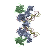
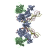
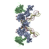
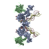
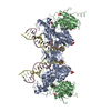
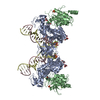
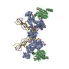
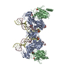
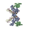
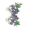
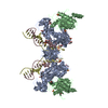
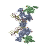
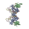
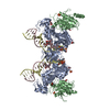
- PDB-3oyc: Crystal structure of the Prototype Foamy Virus (PFV) intasome in ... -
+ Open data

- Basic information
| Entry | Database: PDB / ID: 3oyc | ||||||
|---|---|---|---|---|---|---|---|
| Title | Crystal structure of the Prototype Foamy Virus (PFV) intasome in complex with magnesium and the INSTI PICA | ||||||
 Components Components |
| ||||||
 Keywords Keywords | RECOMBINATION / VIRAL PROTEIN/DNA / PROTEIN-DNA COMPLEX / TETRAMER / DNA INTEGRATION / ENDONUCLEASE / METAL-BINDING / MULTIFUNCTIONAL ENZYME / NUCLEASE / NUCLEOTIDYLTRANSFERASE / NUCLEUS / TRANSFERASE / VIRAL NUCLEOPROTEIN / VIRION / DNA-BINDING / ZINC BINDING / HHCC MOTIF / VIRAL PROTEIN / INHIBITOR / DNA-BINDING PROTEIN-DNA complex / VIRAL PROTEIN-DNA complex | ||||||
| Function / homology |  Function and homology information Function and homology informationHydrolases; Acting on peptide bonds (peptidases); Aspartic endopeptidases / ribonuclease H / DNA integration / viral genome integration into host DNA / establishment of integrated proviral latency / RNA-directed DNA polymerase / virion component / viral penetration into host nucleus / RNA-directed DNA polymerase activity / RNA-DNA hybrid ribonuclease activity ...Hydrolases; Acting on peptide bonds (peptidases); Aspartic endopeptidases / ribonuclease H / DNA integration / viral genome integration into host DNA / establishment of integrated proviral latency / RNA-directed DNA polymerase / virion component / viral penetration into host nucleus / RNA-directed DNA polymerase activity / RNA-DNA hybrid ribonuclease activity / Transferases; Transferring phosphorus-containing groups; Nucleotidyltransferases / host cell / DNA recombination / DNA-directed DNA polymerase / aspartic-type endopeptidase activity / Hydrolases; Acting on ester bonds / host cell cytoplasm / DNA-directed DNA polymerase activity / symbiont entry into host cell / host cell nucleus / proteolysis / RNA binding / metal ion binding Similarity search - Function | ||||||
| Biological species |  Human spumaretrovirus Human spumaretrovirus | ||||||
| Method |  X-RAY DIFFRACTION / SYNCHROTRON / MOLECULAR REPLACEMENT / Resolution: 2.66 Å X-RAY DIFFRACTION / SYNCHROTRON / MOLECULAR REPLACEMENT / Resolution: 2.66 Å | ||||||
 Authors Authors | Hare, S. / Cherepanov, P. | ||||||
 Citation Citation | Journal: Proc.Natl.Acad.Sci.USA / Year: 2010 Title: Molecular mechanisms of retroviral integrase inhibition and the evolution of viral resistance. Authors: Hare, S. / Vos, A.M. / Clayton, R.F. / Thuring, J.W. / Cummings, M.D. / Cherepanov, P. | ||||||
| History |
|
- Structure visualization
| Structure viewer | Molecule: MolmilJmol/JSmol |
|---|
- Downloads & links
-Download
| PDBx/mmCIF format | 3oyc.cif.gz | 156.6 KB | Display | PDBx/mmCIF format |
|---|---|---|---|---|
| PDB format | pdb3oyc.ent.gz | 116.1 KB | Display | PDB format |
| PDBx/mmJSON format | 3oyc.json.gz | Tree view | PDBx/mmJSON format | |
| Others |  Other downloads Other downloads |
-Validation report
| Arichive directory | https://data.pdbj.org/pub/pdb/validation_reports/oy/3oycftp://data.pdbj.org/pub/pdb/validation_reports/oy/3oyc | HTTPS FTP |
|---|
-Related structure data
| Related structure data |  3oyaC 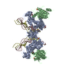 3oybSC 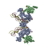 3oydC  3oyeC 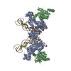 3oyfC 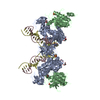 3oygC 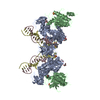 3oyhC  3oyiC 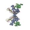 3oyjC 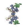 3oykC 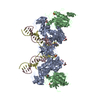 3oylC 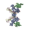 3oymC 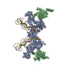 3oynC C: citing same article ( S: Starting model for refinement |
|---|---|
| Similar structure data |


-Links
 PDBj
PDBj


- Assembly
| Deposited unit | 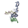
| ||||||||
|---|---|---|---|---|---|---|---|---|---|
| 1 | 
| ||||||||
| Unit cell |
|
-Components
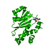
-Protein , 1 types, 2 molecules AB
| #1: Protein | Mass: 44456.695 Da / Num. of mol.: 2 Source method: isolated from a genetically manipulated source Source: (gene. exp.) Human spumaretrovirus / Strain: HSRV2 / Gene: POL / Plasmid: pSSH6P-PFV-INFL / Production host:  |
|---|
-DNA chain , 2 types, 2 molecules CD
| #2: DNA chain | Mass: 5834.794 Da / Num. of mol.: 1 / Source method: obtained synthetically / Details: Donor DNA non-transferred strand |
|---|---|
| #3: DNA chain | Mass: 5195.399 Da / Num. of mol.: 1 / Source method: obtained synthetically / Details: Donor DNA transferred strand |
-Non-polymers , 7 types, 256 molecules 


| #4: Chemical | ChemComp-ZN /  Mass: 65.409 Da / Num. of mol.: 1 / Source method: obtained synthetically / Formula: Zn Mass: 65.409 Da / Num. of mol.: 1 / Source method: obtained synthetically / Formula: Zn | ||||||||||
|---|---|---|---|---|---|---|---|---|---|---|---|
| #5: Chemical |  Mass: 96.063 Da / Num. of mol.: 2 / Source method: obtained synthetically / Formula: SO4 Mass: 96.063 Da / Num. of mol.: 2 / Source method: obtained synthetically / Formula: SO4#6: Chemical | ChemComp-GOL /  Mass: 92.094 Da / Num. of mol.: 4 / Source method: obtained synthetically / Formula: C3H8O3 Mass: 92.094 Da / Num. of mol.: 4 / Source method: obtained synthetically / Formula: C3H8O3#7: Chemical | ChemComp-NH4 / |  Mass: 18.038 Da / Num. of mol.: 1 / Source method: obtained synthetically / Formula: H4N Mass: 18.038 Da / Num. of mol.: 1 / Source method: obtained synthetically / Formula: H4N#8: Chemical |  Mass: 24.305 Da / Num. of mol.: 2 / Source method: obtained synthetically / Formula: Mg Mass: 24.305 Da / Num. of mol.: 2 / Source method: obtained synthetically / Formula: Mg#9: Chemical | ChemComp-ZZW / |  Mass: 335.332 Da / Num. of mol.: 1 / Source method: obtained synthetically / Formula: C19H14FN3O2 Mass: 335.332 Da / Num. of mol.: 1 / Source method: obtained synthetically / Formula: C19H14FN3O2#10: Water | ChemComp-HOH / | Mass: 18.015 Da / Num. of mol.: 245 / Source method: isolated from a natural source / Formula: H2O |
-Experimental details
-Experiment
| Experiment | Method: X-RAY DIFFRACTION / Number of used crystals: 1 |
|---|
- Sample preparation
| Crystal | Density Matthews: 3.96 Å3/Da / Density % sol: 68.95 % |
|---|---|
| Crystal grow | Temperature: 291 K / Method: vapor diffusion, hanging drop / pH: 6.5 Details: 1.35 M ammonium sulfate, 25% (v/v) glycerol, 4.8% (v/v) 1,6-hexanediol, 50 mM Mes-NaOH, 1mM EDTA, pH 6.5, vapor diffusion, hanging drop, temperature 291K |
-Data collection
| Diffraction | Mean temperature: 100 K | ||||||||||||||||||||||||||||||||||||||||||||||||||||||||||||||||||||||||||||||||||||||||
|---|---|---|---|---|---|---|---|---|---|---|---|---|---|---|---|---|---|---|---|---|---|---|---|---|---|---|---|---|---|---|---|---|---|---|---|---|---|---|---|---|---|---|---|---|---|---|---|---|---|---|---|---|---|---|---|---|---|---|---|---|---|---|---|---|---|---|---|---|---|---|---|---|---|---|---|---|---|---|---|---|---|---|---|---|---|---|---|---|---|
| Diffraction source | Source: SYNCHROTRON / Site: SLS  / Beamline: X06DA / Wavelength: 1 Å / Beamline: X06DA / Wavelength: 1 Å | ||||||||||||||||||||||||||||||||||||||||||||||||||||||||||||||||||||||||||||||||||||||||
| Detector | Type: MARMOSAIC 225 mm CCD / Detector: CCD / Date: Sep 18, 2010 | ||||||||||||||||||||||||||||||||||||||||||||||||||||||||||||||||||||||||||||||||||||||||
| Radiation | Monochromator: Bartels Monochromator Si(111) / Protocol: SINGLE WAVELENGTH / Monochromatic (M) / Laue (L): M / Scattering type: x-ray | ||||||||||||||||||||||||||||||||||||||||||||||||||||||||||||||||||||||||||||||||||||||||
| Radiation wavelength | Wavelength: 1 Å / Relative weight: 1 | ||||||||||||||||||||||||||||||||||||||||||||||||||||||||||||||||||||||||||||||||||||||||
| Reflection | Resolution: 2.66→38.796 Å / Num. obs: 46307 / % possible obs: 99.6 % / Observed criterion σ(F): -3 / Observed criterion σ(I): -3 / Redundancy: 3.7 % / Rsym value: 0.077 / Net I/σ(I): 10.4 | ||||||||||||||||||||||||||||||||||||||||||||||||||||||||||||||||||||||||||||||||||||||||
| Reflection shell |
|
- Processing
| Software |
| |||||||||||||||||||||||||||||||||||||||||||||||||||||||||||||||||
|---|---|---|---|---|---|---|---|---|---|---|---|---|---|---|---|---|---|---|---|---|---|---|---|---|---|---|---|---|---|---|---|---|---|---|---|---|---|---|---|---|---|---|---|---|---|---|---|---|---|---|---|---|---|---|---|---|---|---|---|---|---|---|---|---|---|---|
| Refinement | Method to determine structure: MOLECULAR REPLACEMENT Starting model: PDB entry 3OYB Resolution: 2.66→38.8 Å / Cor.coef. Fo:Fc: 0.946 / Cor.coef. Fo:Fc free: 0.931 / WRfactor Rfree: 0.2206 / WRfactor Rwork: 0.1939 / Occupancy max: 1 / Occupancy min: 1 / FOM work R set: 0.852 / SU B: 7.918 / SU ML: 0.162 / SU R Cruickshank DPI: 0.2705 / SU Rfree: 0.2155 / Cross valid method: THROUGHOUT / σ(F): 0 / ESU R Free: 0.216 / Stereochemistry target values: MAXIMUM LIKELIHOOD
| |||||||||||||||||||||||||||||||||||||||||||||||||||||||||||||||||
| Solvent computation | Ion probe radii: 0.8 Å / Shrinkage radii: 0.8 Å / VDW probe radii: 1.4 Å / Solvent model: MASK | |||||||||||||||||||||||||||||||||||||||||||||||||||||||||||||||||
| Displacement parameters | Biso max: 141.87 Å2 / Biso mean: 56.4308 Å2 / Biso min: 25.63 Å2
| |||||||||||||||||||||||||||||||||||||||||||||||||||||||||||||||||
| Refinement step | Cycle: LAST / Resolution: 2.66→38.8 Å
| |||||||||||||||||||||||||||||||||||||||||||||||||||||||||||||||||
| Refine LS restraints |
| |||||||||||||||||||||||||||||||||||||||||||||||||||||||||||||||||
| LS refinement shell | Resolution: 2.66→2.729 Å / Total num. of bins used: 20
|
