Mass: 18.015 Da / Num. of mol.: 48 / Source method: isolated from a natural source / Formula: H2O
Has protein modification
Y
-
Experimental details
-
Experiment
Experiment
Method: X-RAY DIFFRACTION / Number of used crystals: 1
-
Sample preparation
Crystal
Density Matthews: 2.08 Å3/Da / Density % sol: 40.79 % Description: 1cxu was used as a search model for molecular replacement. Solution found in PHASER (Euler angles 310.7, 84.0, 41.7; fractional coordinates -1.272, 0.422, 0.077) was used as a starting ...Description: 1cxu was used as a search model for molecular replacement. Solution found in PHASER (Euler angles 310.7, 84.0, 41.7; fractional coordinates -1.272, 0.422, 0.077) was used as a starting model for density modification in SOLOMON to improve and extend Se-SAD derived phases.
Crystal grow
Temperature: 291 K / Method: vapor diffusion, hanging drop / pH: 6.5 Details: 1.0 M ammonium formate, 0.2 M magnesium cloride, 5 mM dithiotreitol, 0.1 M 2-(N-morpholino)ethanesulfonic acid, pH 6.5, VAPOR DIFFUSION, HANGING DROP, temperature 291K
Resolution: 2.2→45.18 Å / Cor.coef. Fo:Fc: 0.939 / Cor.coef. Fo:Fc free: 0.909 / SU B: 10.771 / SU ML: 0.141 / Cross valid method: THROUGHOUT / ESU R: 0.255 / ESU R Free: 0.216 / Stereochemistry target values: MAXIMUM LIKELIHOOD / Details: HYDROGENS HAVE BEEN ADDED IN THE RIDING POSITIONS
Rfactor
Num. reflection
% reflection
Selection details
Rfree
0.25922
1042
9.8 %
RANDOM
Rwork
0.20905
-
-
-
obs
0.21394
9540
99.2 %
-
Solvent computation
Ion probe radii: 0.8 Å / Shrinkage radii: 0.8 Å / VDW probe radii: 1.2 Å / Solvent model: MASK
Displacement parameters
Biso mean: 32.046 Å2
Baniso -1
Baniso -2
Baniso -3
1-
0.82 Å2
0.41 Å2
0 Å2
2-
-
0.82 Å2
0 Å2
3-
-
-
-1.23 Å2
Refinement step
Cycle: LAST / Resolution: 2.2→45.18 Å
Protein
Nucleic acid
Ligand
Solvent
Total
Num. atoms
1241
0
1
48
1290
Refine LS restraints
Refine-ID
Type
Dev ideal
Dev ideal target
Number
X-RAY DIFFRACTION
f_bond_refined_d
0.01
0.022
1255
X-RAY DIFFRACTION
f_bond_other_d
X-RAY DIFFRACTION
f_angle_refined_deg
1.122
1.969
1712
X-RAY DIFFRACTION
f_angle_other_deg
X-RAY DIFFRACTION
f_dihedral_angle_1_deg
5.838
5
152
X-RAY DIFFRACTION
f_dihedral_angle_2_deg
35.371
23.111
45
X-RAY DIFFRACTION
f_dihedral_angle_3_deg
16.041
15
202
X-RAY DIFFRACTION
f_dihedral_angle_4_deg
16.931
15
4
X-RAY DIFFRACTION
f_chiral_restr
0.073
0.2
201
X-RAY DIFFRACTION
f_gen_planes_refined
0.004
0.02
918
X-RAY DIFFRACTION
f_gen_planes_other
X-RAY DIFFRACTION
f_nbd_refined
0.197
0.2
523
X-RAY DIFFRACTION
f_nbd_other
X-RAY DIFFRACTION
f_nbtor_refined
0.307
0.2
860
X-RAY DIFFRACTION
f_nbtor_other
X-RAY DIFFRACTION
f_xyhbond_nbd_refined
0.114
0.2
60
X-RAY DIFFRACTION
f_xyhbond_nbd_other
X-RAY DIFFRACTION
f_metal_ion_refined
0.099
0.2
1
X-RAY DIFFRACTION
f_metal_ion_other
X-RAY DIFFRACTION
f_symmetry_vdw_refined
0.149
0.2
39
X-RAY DIFFRACTION
f_symmetry_vdw_other
X-RAY DIFFRACTION
f_symmetry_hbond_refined
0.131
0.2
4
X-RAY DIFFRACTION
f_symmetry_hbond_other
X-RAY DIFFRACTION
f_symmetry_metal_ion_refined
X-RAY DIFFRACTION
f_symmetry_metal_ion_other
X-RAY DIFFRACTION
f_mcbond_it
0.558
1.5
791
X-RAY DIFFRACTION
f_mcbond_other
X-RAY DIFFRACTION
f_mcangle_it
1.025
2
1264
X-RAY DIFFRACTION
f_scbond_it
1.746
3
535
X-RAY DIFFRACTION
f_scangle_it
2.472
4.5
448
X-RAY DIFFRACTION
f_rigid_bond_restr
X-RAY DIFFRACTION
f_sphericity_free
X-RAY DIFFRACTION
f_sphericity_bonded
LS refinement shell
Resolution: 2.2→2.257 Å / Total num. of bins used: 20
Rfactor
Num. reflection
% reflection
Rfree
0.223
75
-
Rwork
0.219
683
-
obs
-
-
98.83 %
Refinement TLS params.
Method: refined / Refine-ID: X-RAY DIFFRACTION
ID
L11 (°2)
L12 (°2)
L13 (°2)
L22 (°2)
L23 (°2)
L33 (°2)
S11 (Å °)
S12 (Å °)
S13 (Å °)
S21 (Å °)
S22 (Å °)
S23 (Å °)
S31 (Å °)
S32 (Å °)
S33 (Å °)
T11 (Å2)
T12 (Å2)
T13 (Å2)
T22 (Å2)
T23 (Å2)
T33 (Å2)
Origin x (Å)
Origin y (Å)
Origin z (Å)
1
2.0876
-0.174
-0.1366
1.78
-0.7742
2.6847
0.0058
0.074
-0.16
-0.0405
0.0673
-0.0445
0.2553
0.0082
-0.0731
-0.0593
0.0565
-0.0181
-0.1625
-0.0245
-0.1432
-0.066
19.715
14.154
2
29.9409
13.1846
11.6656
33.5029
-8.8202
17.701
-0.4337
1.7411
0.824
-0.4456
-0.1878
0.5624
-0.2635
1.1541
0.6216
0.013
-0.1786
-0.0727
0.2407
0.0904
-0.1504
-9.21
22.506
-3.052
Refinement TLS group
ID
Refine-ID
Refine TLS-ID
Auth asym-ID
Label asym-ID
Auth seq-ID
Label seq-ID
1
X-RAY DIFFRACTION
1
A
A
119 - 271
14 - 166
2
X-RAY DIFFRACTION
2
A
A
293 - 304
188 - 199
+
About Yorodumi
-
News
-
Feb 9, 2022. New format data for meta-information of EMDB entries
New format data for meta-information of EMDB entries
Version 3 of the EMDB header file is now the official format.
The previous official version 1.9 will be removed from the archive.
In the structure databanks used in Yorodumi, some data are registered as the other names, "COVID-19 virus" and "2019-nCoV". Here are the details of the virus and the list of structure data.
Jan 31, 2019. EMDB accession codes are about to change! (news from PDBe EMDB page)
EMDB accession codes are about to change! (news from PDBe EMDB page)
The allocation of 4 digits for EMDB accession codes will soon come to an end. Whilst these codes will remain in use, new EMDB accession codes will include an additional digit and will expand incrementally as the available range of codes is exhausted. The current 4-digit format prefixed with “EMD-” (i.e. EMD-XXXX) will advance to a 5-digit format (i.e. EMD-XXXXX), and so on. It is currently estimated that the 4-digit codes will be depleted around Spring 2019, at which point the 5-digit format will come into force.
The EM Navigator/Yorodumi systems omit the EMD- prefix.
Related info.:Q: What is EMD? / ID/Accession-code notation in Yorodumi/EM Navigator
Yorodumi is a browser for structure data from EMDB, PDB, SASBDB, etc.
This page is also the successor to EM Navigator detail page, and also detail information page/front-end page for Omokage search.
The word "yorodu" (or yorozu) is an old Japanese word meaning "ten thousand". "mi" (miru) is to see.
Related info.:EMDB / PDB / SASBDB / Comparison of 3 databanks / Yorodumi Search / Aug 31, 2016. New EM Navigator & Yorodumi / Yorodumi Papers / Jmol/JSmol / Function and homology information / Changes in new EM Navigator and Yorodumi
 Movie
Movie Controller

 Yorodumi
 Open data
 Basic information
 Components
 Keywords
 Function and homology information
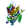
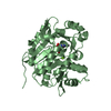
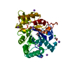
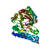
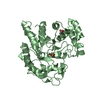
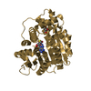
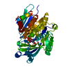
 Human spumaretrovirus
 X-RAY DIFFRACTION /
  Authors
 Citation
 Structure visualization
 Downloads & links
 Other downloads
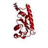

 PDBj
PDBj
 Assembly


 Mass: 24.305 Da / Num. of mol.: 1 / Source method: obtained synthetically / Formula: Mg
 Mass: 18.015 Da / Num. of mol.: 48 / Source method: isolated from a natural source / Formula: H2O
 Sample preparation
 Processing