| 登録情報 | データベース: PDB / ID: 3h0p
|
|---|
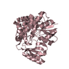
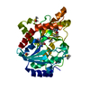
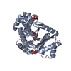
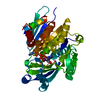
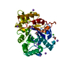
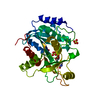
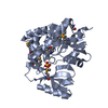
| タイトル | 2.0 Angstrom Crystal Structure of an Acyl Carrier Protein S-malonyltransferase from Salmonella typhimurium. |
|---|
 要素 要素 | S-malonyltransferase |
|---|
 キーワード キーワード | TRANSFERASE / acyl carrier protein / S-malonyltransferase / idp00956 / FabD / Fatty acid biosynthesis / Lipid synthesis / Structural Genomics / Center for Structural Genomics of Infectious Diseases / CSGID |
|---|
| 機能・相同性 |  機能・相同性情報 機能・相同性情報
[acyl-carrier-protein] S-malonyltransferase / [acyl-carrier-protein] S-malonyltransferase activity / fatty acid biosynthetic process / cytosol類似検索 - 分子機能 Malonyl CoA-acyl carrier protein transacylase / Malonyl CoA-acyl carrier protein transacylase, FabD-type / : / Malonyl-CoA ACP transacylase, ACP-binding / Malonyl-Coenzyme A Acyl Carrier Protein, domain 2 / Malonyl-Coenzyme A Acyl Carrier Protein; domain 2 / Malonyl-CoA ACP transacylase, ACP-binding / Acyl transferase / Acyl transferase domain / Acyl transferase domain in polyketide synthase (PKS) enzymes. ...Malonyl CoA-acyl carrier protein transacylase / Malonyl CoA-acyl carrier protein transacylase, FabD-type / : / Malonyl-CoA ACP transacylase, ACP-binding / Malonyl-Coenzyme A Acyl Carrier Protein, domain 2 / Malonyl-Coenzyme A Acyl Carrier Protein; domain 2 / Malonyl-CoA ACP transacylase, ACP-binding / Acyl transferase / Acyl transferase domain / Acyl transferase domain in polyketide synthase (PKS) enzymes. / Acyl transferase domain superfamily / Acyl transferase/acyl hydrolase/lysophospholipase / Alpha-Beta Plaits / 2-Layer Sandwich / 3-Layer(aba) Sandwich / Alpha Beta類似検索 - ドメイン・相同性 Malonyl CoA-acyl carrier protein transacylase類似検索 - 構成要素 |
|---|
| 生物種 |  Salmonella typhimurium (サルモネラ菌) Salmonella typhimurium (サルモネラ菌) |
|---|
| 手法 |  X線回折 / シンクロトロン / 分子置換 / 解像度: 2 Å X線回折 / シンクロトロン / 分子置換 / 解像度: 2 Å |
|---|
 データ登録者 データ登録者 | Minasov, G. / Wawrzak, Z. / Skarina, T. / Onopriyenko, O. / Peterson, S.N. / Savchenko, A. / Anderson, W.F. / Center for Structural Genomics of Infectious Diseases (CSGID) / Center for Structural Genomics of Infectious Diseases (CSGID) |
|---|
 引用 引用 | ジャーナル: TO BE PUBLISHED
タイトル: 2.0 Angstrom Crystal Structure of an Acyl Carrier Protein S-malonyltransferase from Salmonella typhimurium.
著者: Minasov, G. / Wawrzak, Z. / Skarina, T. / Onopriyenko, O. / Peterson, S.N. / Savchenko, A. / Anderson, W.F. / Center for Structural Genomics of Infectious Diseases (CSGID) |
|---|
| 履歴 | | 登録 | 2009年4月10日 | 登録サイト: RCSB / 処理サイト: RCSB |
|---|
| 改定 1.0 | 2009年4月21日 | Provider: repository / タイプ: Initial release |
|---|
| 改定 1.1 | 2011年7月13日 | Group: Advisory / Version format compliance |
|---|
| 改定 1.2 | 2017年11月1日 | Group: Refinement description / カテゴリ: software / Item: _software.name |
|---|
| 改定 1.3 | 2023年9月6日 | Group: Data collection / Database references ...Data collection / Database references / Derived calculations / Refinement description
カテゴリ: chem_comp_atom / chem_comp_bond ...chem_comp_atom / chem_comp_bond / database_2 / pdbx_initial_refinement_model / struct_conn / struct_ref_seq_dif / struct_site
Item: _database_2.pdbx_DOI / _database_2.pdbx_database_accession ..._database_2.pdbx_DOI / _database_2.pdbx_database_accession / _struct_conn.pdbx_leaving_atom_flag / _struct_ref_seq_dif.details / _struct_site.pdbx_auth_asym_id / _struct_site.pdbx_auth_comp_id / _struct_site.pdbx_auth_seq_id |
|---|
| 改定 1.4 | 2023年11月22日 | Group: Data collection / カテゴリ: chem_comp_atom / chem_comp_bond / Item: _chem_comp_atom.atom_id / _chem_comp_bond.atom_id_2 |
|---|
| 改定 1.5 | 2024年11月6日 | Group: Structure summary
カテゴリ: pdbx_entry_details / pdbx_modification_feature |
|---|
|
|---|
 ムービー
ムービー コントローラー


 データを開く
 基本情報
基本情報 構造の表示
構造の表示 ダウンロードとリンク
 その他のダウンロード


 PDBj
PDBj
 集合体
集合体


 分子量: 96.063 Da / 分子数: 3 / 由来タイプ: 合成 / 式: SO4
 分子量: 18.015 Da / 分子数: 443 / 由来タイプ: 天然 / 式: H2O
 試料調製
試料調製 / ビームライン: 21-ID-D / 波長: 0.98011 Å
 解析
解析