 Movie
Movie Controller


+ Open data

- Basic information
| Entry | Database: PDB / ID: 3ou1 | ||||||
|---|---|---|---|---|---|---|---|
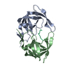
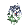
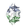
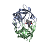
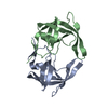
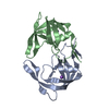
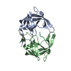
| Title | MDR769 HIV-1 protease complexed with RH/IN hepta-peptide | ||||||
 Components Components |
| ||||||
 Keywords Keywords | HYDROLASE/PEPTIDE / MDR HIV-1 protease / inhibitor / drug resistance / substrate envelope / HIV-1 protease / protease / substrate peptide / HYDROLASE / HYDROLASE-PEPTIDE complex | ||||||
| Function / homology |  Function and homology information Function and homology informationHIV-1 retropepsin / symbiont-mediated activation of host apoptosis / retroviral ribonuclease H / exoribonuclease H / exoribonuclease H activity / host multivesicular body / DNA integration / viral genome integration into host DNA / RNA-directed DNA polymerase / establishment of integrated proviral latency ...HIV-1 retropepsin / symbiont-mediated activation of host apoptosis / retroviral ribonuclease H / exoribonuclease H / exoribonuclease H activity / host multivesicular body / DNA integration / viral genome integration into host DNA / RNA-directed DNA polymerase / establishment of integrated proviral latency / viral penetration into host nucleus / RNA stem-loop binding / RNA-directed DNA polymerase activity / RNA-DNA hybrid ribonuclease activity / Transferases; Transferring phosphorus-containing groups; Nucleotidyltransferases / host cell / viral nucleocapsid / endonuclease activity / DNA recombination / DNA-directed DNA polymerase / aspartic-type endopeptidase activity / Hydrolases; Acting on ester bonds / DNA-directed DNA polymerase activity / symbiont-mediated suppression of host gene expression / viral translational frameshifting / lipid binding / symbiont entry into host cell / host cell nucleus / host cell plasma membrane / virion membrane / structural molecule activity / proteolysis / DNA binding / zinc ion binding / membrane Similarity search - Function | ||||||
| Biological species |   Human immunodeficiency virus 1 Human immunodeficiency virus 1 | ||||||
| Method |  X-RAY DIFFRACTION / SYNCHROTRON / MOLECULAR REPLACEMENT / Resolution: 1.8 Å X-RAY DIFFRACTION / SYNCHROTRON / MOLECULAR REPLACEMENT / Resolution: 1.8 Å | ||||||
 Authors Authors | Liu, Z. / Wang, Y. / Brunzelle, J. / Kovari, I.A. / Kovari, L.C. | ||||||
 Citation Citation | Journal: Protein J. / Year: 2011 Title: Nine Crystal Structures Determine the Substrate Envelope of the MDR HIV-1 Protease. Authors: Liu, Z. / Wang, Y. / Brunzelle, J. / Kovari, I.A. / Kovari, L.C. | ||||||
| History |
|
- Structure visualization
| Structure viewer | Molecule: MolmilJmol/JSmol |
|---|
- Downloads & links
-Download
| PDBx/mmCIF format | 3ou1.cif.gz | 56.8 KB | Display | PDBx/mmCIF format |
|---|---|---|---|---|
| PDB format | pdb3ou1.ent.gz | 41.5 KB | Display | PDB format |
| PDBx/mmJSON format | 3ou1.json.gz | Tree view | PDBx/mmJSON format | |
| Others |  Other downloads Other downloads |
-Validation report
| Summary document | 3ou1_validation.pdf.gz | 440.1 KB | Display | wwPDB validaton report |
|---|---|---|---|---|
| Full document | 3ou1_full_validation.pdf.gz | 445.1 KB | Display | |
| Data in XML | 3ou1_validation.xml.gz | 13.3 KB | Display | |
| Data in CIF | 3ou1_validation.cif.gz | 18.6 KB | Display | |
| Arichive directory | https://data.pdbj.org/pub/pdb/validation_reports/ou/3ou1ftp://data.pdbj.org/pub/pdb/validation_reports/ou/3ou1 | HTTPS FTP |
-Related structure data
| Related structure data |  3otsC  3otyC  3ou3C  3ou4C  3ouaC 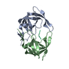 3oubC  3oucC  3oudC C: citing same article ( |
|---|---|
| Similar structure data |


-Links
 PDBj
PDBj

- Assembly
| Deposited unit | 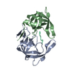
| ||||||||
|---|---|---|---|---|---|---|---|---|---|
| 1 |
| ||||||||
| Unit cell |
| ||||||||
| Details | HIV-1 protease dimer binds with RH/IN substrate peptide |
-Components
| #1: Protein | Mass: 10769.635 Da / Num. of mol.: 1 Source method: isolated from a genetically manipulated source Source: (gene. exp.) Human immunodeficiency virus 1 / Gene: pol / Production host:  |
|---|---|
| #2: Protein | Mass: 10771.607 Da / Num. of mol.: 1 Source method: isolated from a genetically manipulated source Source: (gene. exp.) Human immunodeficiency virus 1 / Gene: pol / Production host: |
| #3: Protein/peptide | Mass: 791.955 Da / Num. of mol.: 1 / Source method: obtained synthetically / Details: This sequence occurs naturally in HIV-1 / Source: (synth.) Human immunodeficiency virus 1 / References: UniProt: Q9YV20, UniProt: P35963*PLUS |
| #4: Water | ChemComp-HOH /  Mass: 18.015 Da / Num. of mol.: 238 / Source method: isolated from a natural source / Formula: H2O Mass: 18.015 Da / Num. of mol.: 238 / Source method: isolated from a natural source / Formula: H2O |
-Experimental details
-Experiment
| Experiment | Method: X-RAY DIFFRACTION / Number of used crystals: 1 |
|---|
- Sample preparation
| Crystal | Density Matthews: 2.37 Å3/Da / Density % sol: 48.1 % |
|---|---|
| Crystal grow | Temperature: 298 K / Method: vapor diffusion, hanging drop / pH: 5.8 Details: 0.8M NaCl 01 M MES , pH 5.8, VAPOR DIFFUSION, HANGING DROP, temperature 298K |
-Data collection
| Diffraction | Mean temperature: 173 K | ||||||||||||||||||||||
|---|---|---|---|---|---|---|---|---|---|---|---|---|---|---|---|---|---|---|---|---|---|---|---|
| Diffraction source | Source: SYNCHROTRON / Site: APS  / Beamline: 21-ID-D / Wavelength: 1.0332 Å / Beamline: 21-ID-D / Wavelength: 1.0332 Å | ||||||||||||||||||||||
| Detector | Type: MARMOSAIC 300 mm CCD / Detector: CCD / Date: Aug 9, 2008 | ||||||||||||||||||||||
| Radiation | Monochromator: kohzu / Protocol: SINGLE WAVELENGTH / Monochromatic (M) / Laue (L): M / Scattering type: x-ray | ||||||||||||||||||||||
| Radiation wavelength | Wavelength: 1.0332 Å / Relative weight: 1 | ||||||||||||||||||||||
| Reflection | Resolution: 1.8→45 Å / Num. all: 19214 / Num. obs: 19157 / % possible obs: 99.7 % / Observed criterion σ(F): 1 / Observed criterion σ(I): 1 | ||||||||||||||||||||||
| Reflection shell |
|
- Processing
| Software |
| ||||||||||||||||||||||||||||||||||||||||||||||||||||||||||||||||||||||||||||||||||||||||||
|---|---|---|---|---|---|---|---|---|---|---|---|---|---|---|---|---|---|---|---|---|---|---|---|---|---|---|---|---|---|---|---|---|---|---|---|---|---|---|---|---|---|---|---|---|---|---|---|---|---|---|---|---|---|---|---|---|---|---|---|---|---|---|---|---|---|---|---|---|---|---|---|---|---|---|---|---|---|---|---|---|---|---|---|---|---|---|---|---|---|---|---|
| Refinement | Method to determine structure: MOLECULAR REPLACEMENT / Resolution: 1.8→41.34 Å / Cor.coef. Fo:Fc: 0.952 / Cor.coef. Fo:Fc free: 0.922 / SU B: 2.75 / SU ML: 0.088 / Cross valid method: THROUGHOUT / σ(F): 0 / ESU R Free: 0.137 / Stereochemistry target values: MAXIMUM LIKELIHOOD / Details: HYDROGENS HAVE BEEN ADDED IN THE RIDING POSITIONS
| ||||||||||||||||||||||||||||||||||||||||||||||||||||||||||||||||||||||||||||||||||||||||||
| Solvent computation | Ion probe radii: 0.8 Å / Shrinkage radii: 0.8 Å / VDW probe radii: 1.2 Å / Solvent model: MASK | ||||||||||||||||||||||||||||||||||||||||||||||||||||||||||||||||||||||||||||||||||||||||||
| Displacement parameters | Biso mean: 23.095 Å2
| ||||||||||||||||||||||||||||||||||||||||||||||||||||||||||||||||||||||||||||||||||||||||||
| Refinement step | Cycle: LAST / Resolution: 1.8→41.34 Å
| ||||||||||||||||||||||||||||||||||||||||||||||||||||||||||||||||||||||||||||||||||||||||||
| Refine LS restraints |
| ||||||||||||||||||||||||||||||||||||||||||||||||||||||||||||||||||||||||||||||||||||||||||
| LS refinement shell | Resolution: 1.8→1.847 Å / Total num. of bins used: 20
|
