 ムービー
ムービー コントローラー


+ データを開く

- 基本情報
基本情報
| 登録情報 | データベース: PDB / ID: 3oov | ||||||
|---|---|---|---|---|---|---|---|
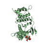
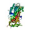
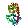
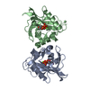
| タイトル | Crystal structure of a methyl-accepting chemotaxis protein, residues 122 to 287 | ||||||
 要素 要素 | Methyl-accepting chemotaxis protein, putative | ||||||
 キーワード キーワード | SIGNALING PROTEIN / Structural Genomics / PSI-2 / Protein Structure Initiative / Midwest Center for Structural Genomics / MCSG | ||||||
| 機能・相同性 |  機能・相同性情報 機能・相同性情報chemotaxis / transmembrane signaling receptor activity / signal transduction / plasma membrane 類似検索 - 分子機能 | ||||||
| 生物種 |  Geobacter sulfurreducens (バクテリア) Geobacter sulfurreducens (バクテリア) | ||||||
| 手法 |  X線回折 / シンクロトロン / 単波長異常分散 / 解像度: 2.2 Å X線回折 / シンクロトロン / 単波長異常分散 / 解像度: 2.2 Å | ||||||
 データ登録者 データ登録者 | Joachimiak, A. / Duke, N.E.C. / Hatzos-Skintges, C. / Mulligan, R. / Clancy, S. / Midwest Center for Structural Genomics (MCSG) | ||||||
 引用 引用 | ジャーナル: To be Published タイトル: Crystal structure of a methyl-accepting chemotaxis protein, residues 122 to 287 著者: Joachimiak, A. / Duke, N.E.C. / Hatzos-Skintges, C. / Mulligan, R. / Clancy, S. | ||||||
| 履歴 |
|
- 構造の表示
構造の表示
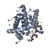
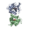
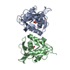
| 構造ビューア | 分子: MolmilJmol/JSmol |
|---|
- ダウンロードとリンク
-ダウンロード
| PDBx/mmCIF形式 | 3oov.cif.gz | 78.5 KB | 表示 | PDBx/mmCIF形式 |
|---|---|---|---|---|
| PDB形式 | pdb3oov.ent.gz | 59.8 KB | 表示 | PDB形式 |
| PDBx/mmJSON形式 | 3oov.json.gz | ツリー表示 | PDBx/mmJSON形式 | |
| その他 |  その他のダウンロード その他のダウンロード |
-検証レポート
| アーカイブディレクトリ | https://data.pdbj.org/pub/pdb/validation_reports/oo/3oovftp://data.pdbj.org/pub/pdb/validation_reports/oo/3oov | HTTPS FTP |
|---|
-関連構造データ
| 類似構造データ | |
|---|---|
| その他のデータベース |


-リンク
 PDBj
PDBj


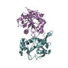
- 集合体
集合体
| 登録構造単位 | 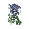
| ||||||||
|---|---|---|---|---|---|---|---|---|---|
| 1 |
| ||||||||
| 2 | 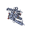
| ||||||||
| 3 | 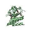
| ||||||||
| 単位格子 |
| ||||||||
| Components on special symmetry positions |
|
-要素
| #1: タンパク質 | 分子量: 18637.951 Da / 分子数: 2 / 断片: residues 122-287 / 由来タイプ: 組換発現 由来: (組換発現) Geobacter sulfurreducens (バクテリア)株: PCA / 遺伝子: GSU1704 / プラスミド: pMCSG7 / 発現宿主: #2: 化合物 | ChemComp-GOL /   分子量: 92.094 Da / 分子数: 4 / 由来タイプ: 合成 / 式: C3H8O3 分子量: 92.094 Da / 分子数: 4 / 由来タイプ: 合成 / 式: C3H8O3#3: 水 | ChemComp-HOH / |  分子量: 18.015 Da / 分子数: 145 / 由来タイプ: 天然 / 式: H2O 分子量: 18.015 Da / 分子数: 145 / 由来タイプ: 天然 / 式: H2OHas protein modification | Y | |
|---|
-実験情報
-実験
| 実験 | 手法: X線回折 / 使用した結晶の数: 1 |
|---|
- 試料調製
試料調製
| 結晶 | マシュー密度: 2.54 Å3/Da / 溶媒含有率: 51.61 % |
|---|---|
| 結晶化 | 温度: 289 K / 手法: 蒸気拡散法, シッティングドロップ法 / pH: 4.2 詳細: 10% w/v PEG 3000, 0.10M phosphate-citrate, pH4.2, 0.20M NaCl. (Wizard II, #36) cryoprotectant: included all of above, in addition to 25% glycerol, VAPOR DIFFUSION, SITTING DROP, temperature 289K |
-データ収集
| 回折 | 平均測定温度: 100 K |
|---|---|
| 放射光源 | 由来: シンクロトロン / サイト: APS  / ビームライン: 19-ID / 波長: 0.97911 Å / ビームライン: 19-ID / 波長: 0.97911 Å |
| 検出器 | タイプ: ADSC QUANTUM 315r / 検出器: CCD / 日付: 2009年11月9日 |
| 放射 | モノクロメーター: double crystal Si(111) monochromator, focussing mirror プロトコル: SINGLE WAVELENGTH / 単色(M)・ラウエ(L): M / 散乱光タイプ: x-ray |
| 放射波長 | 波長: 0.97911 Å / 相対比: 1 |
| 反射 | 解像度: 2.2→60.52 Å / Num. all: 21188 / % possible obs: 99.4 % / Observed criterion σ(I): 3 / 冗長度: 4.1 % |
- 解析
解析
| ソフトウェア |
| |||||||||||||||||||||||||||||||||||||||||||||||||||||||||||||||||
|---|---|---|---|---|---|---|---|---|---|---|---|---|---|---|---|---|---|---|---|---|---|---|---|---|---|---|---|---|---|---|---|---|---|---|---|---|---|---|---|---|---|---|---|---|---|---|---|---|---|---|---|---|---|---|---|---|---|---|---|---|---|---|---|---|---|---|
| 精密化 | 構造決定の手法: 単波長異常分散 / 解像度: 2.2→60.52 Å / Cor.coef. Fo:Fc: 0.926 / Cor.coef. Fo:Fc free: 0.891 / SU B: 6.397 / SU ML: 0.164 / 交差検証法: THROUGHOUT / ESU R Free: 0.251 / 立体化学のターゲット値: MAXIMUM LIKELIHOOD / 詳細: HYDROGENS HAVE BEEN ADDED IN THE RIDING POSITIONS
| |||||||||||||||||||||||||||||||||||||||||||||||||||||||||||||||||
| 溶媒の処理 | イオンプローブ半径: 0.8 Å / 減衰半径: 0.8 Å / VDWプローブ半径: 1.4 Å / 溶媒モデル: MASK | |||||||||||||||||||||||||||||||||||||||||||||||||||||||||||||||||
| 原子変位パラメータ | Biso mean: 28.315 Å2
| |||||||||||||||||||||||||||||||||||||||||||||||||||||||||||||||||
| 精密化ステップ | サイクル: LAST / 解像度: 2.2→60.52 Å
| |||||||||||||||||||||||||||||||||||||||||||||||||||||||||||||||||
| 拘束条件 |
| |||||||||||||||||||||||||||||||||||||||||||||||||||||||||||||||||
| LS精密化 シェル | 解像度: 2.2→2.257 Å / Total num. of bins used: 20
|
