[English] 日本語
Yorodumi
- PDB-3oo6: Crystal structures and biochemical characterization of the bacter... -

+
Open data


ID or keywords:

Loading...

-
Basic information

Entry
Database: PDB / ID: 3oo6
Title
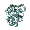
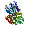
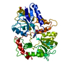
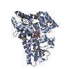
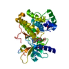
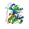
Crystal structures and biochemical characterization of the bacterial solute receptor AcbH reveal an unprecedented exclusive substrate preference for b-D-galactopyranose
ComponentsABC transporter binding protein AcbH
KeywordsSUGAR BINDING PROTEIN / class 2 SBP fold / ABC transporter extracellular solute binding protein / D-galactose Binding
Function / homology
Function and homology information


hydrolase activity, acting on glycosyl bonds / xylan catabolic process / maltose binding / maltose transport / maltodextrin transmembrane transport / ATP-binding cassette (ABC) transporter complex, substrate-binding subunit-containing
Similarity search - Function
Bacterial extracellular solute-binding protein / Bacterial extracellular solute-binding protein / Twin arginine translocation (Tat) signal profile. / Twin-arginine translocation pathway, signal sequence / Periplasmic binding protein-like II / D-Maltodextrin-Binding Protein; domain 2 / Prokaryotic membrane lipoprotein lipid attachment site profile. / 3-Layer(aba) Sandwich / Alpha Beta
Similarity search - Domain/homology
beta-D-galactopyranose / ABC transporter binding protein AcbH
Similarity search - Component
Biological speciesActinoplanes (bacteria)
MethodX-RAY DIFFRACTION / SYNCHROTRON / MOLECULAR REPLACEMENT / Resolution: 2.15 Å
AuthorsVahedi-Faridi, A. / Bulut, H. / Licht, A.
CitationJournal: J.Mol.Biol. / Year: 2011
Title: Crystal structures of the bacterial solute receptor AcbH displaying an exclusive substrate preference for beta-D-galactopyranose
Authors: Licht, A. / Bulut, H. / Scheffel, F. / Daumke, O. / Wehmeier, U.F. / Saenger, W. / Schneider, E. / Vahedi-Faridi, A.
History
DepositionAug 30, 2010Deposition site: RCSB / Processing site: PDBJ
Revision 1.0Dec 29, 2010Provider: repository / Type: Initial release
Revision 1.1Jul 13, 2011Group: Version format compliance
Revision 1.2Mar 26, 2014Group: Database references
Revision 1.3Jul 29, 2020Group: Data collection / Database references ...Data collection / Database references / Derived calculations / Structure summary
Category: chem_comp / entity ...chem_comp / entity / pdbx_chem_comp_identifier / pdbx_entity_nonpoly / struct_ref_seq_dif / struct_site / struct_site_gen
Item: _chem_comp.name / _chem_comp.type ..._chem_comp.name / _chem_comp.type / _entity.pdbx_description / _pdbx_entity_nonpoly.name / _struct_ref_seq_dif.details
Description: Carbohydrate remediation / Provider: repository / Type: Remediation
Revision 1.4Mar 20, 2024Group: Data collection / Database references / Structure summary
Category: chem_comp / chem_comp_atom ...chem_comp / chem_comp_atom / chem_comp_bond / database_2
Item: _chem_comp.pdbx_synonyms / _database_2.pdbx_DOI / _database_2.pdbx_database_accession
Revision 1.5Apr 3, 2024Group: Refinement description / Category: pdbx_initial_refinement_model

-
Structure visualization

Structure viewerMolecule:
MolmilJmol/JSmol

Downloads & links

-
Assembly

Deposited unit
A: ABC transporter binding protein AcbH
B: ABC transporter binding protein AcbH
hetero molecules


Theoretical massNumber of molelcules
Total (without water)92,2836
Polymers91,7312
Non-polymers5524
Water11,836657
1
A: ABC transporter binding protein AcbH
hetero molecules


Theoretical massNumber of molelcules
Total (without water)46,0462
Polymers45,8651
Non-polymers1801
Water181
TypeNameSymmetry operationNumber
identity operation1_555x,y,z1
MethodPISA
2
B: ABC transporter binding protein AcbH
hetero molecules


Theoretical massNumber of molelcules
Total (without water)46,2384
Polymers45,8651
Non-polymers3723
Water181
TypeNameSymmetry operationNumber
identity operation1_555x,y,z1
MethodPISA
Unit cell
Length a, b, c (Å)47.835, 98.812, 95.732
Angle α, β, γ (deg.)90.000, 95.040, 90.000
Int Tables number4
Space group name H-MP1211

-
Components

#1: Protein ABC transporter binding protein AcbH


Mass: 45865.352 Da / Num. of mol.: 2 / Fragment: residue in UNP 39-433 / Mutation: T10M
Source method: isolated from a genetically manipulated source
Source: (gene. exp.) Actinoplanes (bacteria) / Strain: SE50/110 / Gene: AcbH / Plasmid: pAL8, pET15b / Production host: Escherichia coli (E. coli) / Strain (production host): Rosetta2 / References: UniProt: Q27GR2
#2: Sugar ChemComp-GAL / beta-D-galactopyranose / beta-D-galactose / D-galactose / galactose


Type: D-saccharide, beta linking / Mass: 180.156 Da / Num. of mol.: 2
Source method: isolated from a genetically manipulated source
Formula: C6H12O6
IdentifierTypeProgram
DGalpbCONDENSED IUPAC CARBOHYDRATE SYMBOLGMML 1.0
b-D-galactopyranoseCOMMON NAMEGMML 1.0
b-D-GalpIUPAC CARBOHYDRATE SYMBOLPDB-CARE 1.0
GalSNFG CARBOHYDRATE SYMBOLGMML 1.0
#3: Chemical ChemComp-SO4 / SULFATE ION


Mass: 96.063 Da / Num. of mol.: 2 / Source method: obtained synthetically / Formula: SO4
#4: Water ChemComp-HOH / water


Mass: 18.015 Da / Num. of mol.: 657 / Source method: isolated from a natural source / Formula: H2O

-
Experimental details

-
Experiment

ExperimentMethod: X-RAY DIFFRACTION / Number of used crystals: 1

-
Sample preparation

CrystalDensity Matthews: 2.46 Å3/Da / Density % sol: 49.94 % / Mosaicity: 0.419 °
Crystal growTemperature: 291 K / Method: vapor diffusion, hanging drop / pH: 4.5
Details: 2.2M (NH4)2SO4, 100mM citric acid, pH 4.5, VAPOR DIFFUSION, HANGING DROP, temperature 291K

-
Data collection

DiffractionMean temperature: 100 K
Diffraction sourceSource: SYNCHROTRON / Site: BESSY / Beamline: 14.2 / Wavelength: 0.91841 Å
DetectorType: ADSC QUANTUM 210 / Detector: CCD / Date: Aug 23, 2007 / Details: mirrors
RadiationMonochromator: Si 111 CHANNEL / Protocol: SINGLE WAVELENGTH / Monochromatic (M) / Laue (L): M / Scattering type: x-ray
Radiation wavelengthWavelength: 0.91841 Å / Relative weight: 1
ReflectionResolution: 2.15→50 Å / Num. obs: 44711 / % possible obs: 93.1 % / Redundancy: 2.9 % / Rmerge(I) obs: 0.084 / Χ2: 0.908 / Net I/σ(I): 6.8
Reflection shell
Resolution (Å)Redundancy (%)Rmerge(I) obsNum. unique allΧ2Diffraction-ID% possible all
2.15-2.232.50.23439540.856183.2
2.23-2.322.80.20841220.776186.9
2.32-2.422.80.19142230.797187.9
2.42-2.552.90.16942890.817189.3
2.55-2.712.90.14843560.88191.3
2.71-2.922.90.11645440.893194.8
2.92-3.212.90.08747491.021198.6
3.21-3.683.10.06448091.086199.8
3.68-4.633.20.0547921.064199.9
4.63-503.20.04548730.789199.2

-
Processing

Software
NameVersionClassificationNB
SCALEPACKdata scaling
REFMACrefinement
PDB_EXTRACT3.1data extraction
ADSCQuantumdata collection
DENZOdata reduction
PHASERphasing
RefinementMethod to determine structure: MOLECULAR REPLACEMENT
Starting model: model from SeMet SAD phasing

Resolution: 2.15→34.31 Å / Cor.coef. Fo:Fc: 0.944 / Cor.coef. Fo:Fc free: 0.903 / WRfactor Rfree: 0.2299 / WRfactor Rwork: 0.1715 / Occupancy max: 1 / Occupancy min: 0.5 / FOM work R set: 0.8094 / SU B: 5.15 / SU ML: 0.137 / SU R Cruickshank DPI: 0.2812 / SU Rfree: 0.2253 / Cross valid method: THROUGHOUT / σ(F): 0 / ESU R Free: 0.225 / Stereochemistry target values: MAXIMUM LIKELIHOOD
Details: HYDROGENS HAVE BEEN ADDED IN THE RIDING POSITIONS U VALUES: REFINED INDIVIDUALLY
RfactorNum. reflection% reflectionSelection details
Rfree0.2509 2235 5 %RANDOM
Rwork0.1877 ---
obs0.1909 44697 92.9 %-
Solvent computationIon probe radii: 0.8 Å / Shrinkage radii: 0.8 Å / VDW probe radii: 1.4 Å / Solvent model: MASK
Displacement parametersBiso max: 47.57 Å2 / Biso mean: 13.203 Å2 / Biso min: 2 Å2
Baniso -1Baniso -2Baniso -3
1--1.72 Å20 Å2-0.43 Å2
2--2.14 Å20 Å2
3----0.49 Å2
Refinement stepCycle: LAST / Resolution: 2.15→34.31 Å
ProteinNucleic acidLigandSolventTotal
Num. atoms6100 0 34 657 6791
Refine LS restraints
Refine-IDTypeDev idealDev ideal targetNumber
X-RAY DIFFRACTIONr_bond_refined_d0.0110.0226308
X-RAY DIFFRACTIONr_angle_refined_deg0.9311.9428592
X-RAY DIFFRACTIONr_dihedral_angle_1_deg8.7765782
X-RAY DIFFRACTIONr_dihedral_angle_2_deg34.62725.411292
X-RAY DIFFRACTIONr_dihedral_angle_3_deg17.86115990
X-RAY DIFFRACTIONr_dihedral_angle_4_deg24.6791514
X-RAY DIFFRACTIONr_chiral_restr0.0830.2928
X-RAY DIFFRACTIONr_gen_planes_refined0.010.0214874
X-RAY DIFFRACTIONr_mcbond_it0.8171.53894
X-RAY DIFFRACTIONr_mcangle_it1.37226258
X-RAY DIFFRACTIONr_scbond_it2.60232414
X-RAY DIFFRACTIONr_scangle_it3.7634.52332
LS refinement shellResolution: 2.152→2.208 Å / Total num. of bins used: 20
RfactorNum. reflection% reflection
Rfree0.293 131 -
Rwork0.199 2648 -
all-2779 -
obs--79.54 %

+
About Yorodumi

-
News

-
Feb 9, 2022. New format data for meta-information of EMDB entries

New format data for meta-information of EMDB entries

  • Version 3 of the EMDB header file is now the official format.
  • The previous official version 1.9 will be removed from the archive.

Related info.:EMDB header

External links:wwPDB to switch to version 3 of the EMDB data model

-
Aug 12, 2020. Covid-19 info

Covid-19 info

URL: https://pdbjlvh1.pdbj.org/emnavi/covid19.php

New page: Covid-19 featured information page in EM Navigator.

Related info.:Covid-19 info / Mar 5, 2020. Novel coronavirus structure data

+
Mar 5, 2020. Novel coronavirus structure data

Novel coronavirus structure data

Related info.:Yorodumi Speices / Aug 12, 2020. Covid-19 info

External links:COVID-19 featured content - PDBj / Molecule of the Month (242):Coronavirus Proteases

+
Jan 31, 2019. EMDB accession codes are about to change! (news from PDBe EMDB page)

EMDB accession codes are about to change! (news from PDBe EMDB page)

  • The allocation of 4 digits for EMDB accession codes will soon come to an end. Whilst these codes will remain in use, new EMDB accession codes will include an additional digit and will expand incrementally as the available range of codes is exhausted. The current 4-digit format prefixed with “EMD-” (i.e. EMD-XXXX) will advance to a 5-digit format (i.e. EMD-XXXXX), and so on. It is currently estimated that the 4-digit codes will be depleted around Spring 2019, at which point the 5-digit format will come into force.
  • The EM Navigator/Yorodumi systems omit the EMD- prefix.

Related info.:Q: What is EMD? / ID/Accession-code notation in Yorodumi/EM Navigator

External links:EMDB Accession Codes are Changing Soon! / Contact to PDBj

+
Jul 12, 2017. Major update of PDB

Major update of PDB

  • wwPDB released updated PDB data conforming to the new PDBx/mmCIF dictionary.
  • This is a major update changing the version number from 4 to 5, and with Remediation, in which all the entries are updated.
  • In this update, many items about electron microscopy experimental information are reorganized (e.g. em_software).
  • Now, EM Navigator and Yorodumi are based on the updated data.

External links:wwPDB Remediation / Enriched Model Files Conforming to OneDep Data Standards Now Available in the PDB FTP Archive

-
Yorodumi

Thousand views of thousand structures

  • Yorodumi is a browser for structure data from EMDB, PDB, SASBDB, etc.
  • This page is also the successor to EM Navigator detail page, and also detail information page/front-end page for Omokage search.
  • The word "yorodu" (or yorozu) is an old Japanese word meaning "ten thousand". "mi" (miru) is to see.

Related info.:EMDB / PDB / SASBDB / Comparison of 3 databanks / Yorodumi Search / Aug 31, 2016. New EM Navigator & Yorodumi / Yorodumi Papers / Jmol/JSmol / Function and homology information / Changes in new EM Navigator and Yorodumi

Read more