 Movie
Movie Controller

[English] 日本語
 Yorodumi
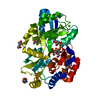
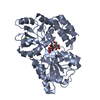
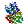
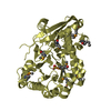
- PDB-4thi: THIAMINASE I FROM BACILLUS THIAMINOLYTICUS WITH COVALENTLY BOUND ... -
+ Open data

- Basic information
| Entry | Database: PDB / ID: 4thi | ||||||
|---|---|---|---|---|---|---|---|
| Title | THIAMINASE I FROM BACILLUS THIAMINOLYTICUS WITH COVALENTLY BOUND 4-AMINO-2,5-DIMETHYLPYRIMIDINE | ||||||
 Components Components | PROTEIN (THIAMINASE I) | ||||||
 Keywords Keywords | TRANSFERASE / THIAMIN DEGRADATION | ||||||
| Function / homology |  Function and homology information Function and homology informationthiamine pyridinylase / thiamine pyridinylase activity / thiamine catabolic process / extracellular region Similarity search - Function | ||||||
| Biological species |  | ||||||
| Method |  X-RAY DIFFRACTION / SYNCHROTRON / MOLECULAR REPLACEMENT / Resolution: 2 Å X-RAY DIFFRACTION / SYNCHROTRON / MOLECULAR REPLACEMENT / Resolution: 2 Å | ||||||
 Authors Authors | Campobasso, N. / Begley, T.P. / Ealick, S.E. | ||||||
 Citation Citation | Journal: Biochemistry / Year: 1998 Title: Crystal structure of thiaminase-I from Bacillus thiaminolyticus at 2.0 A resolution. Authors: Campobasso, N. / Costello, C.A. / Kinsland, C. / Begley, T.P. / Ealick, S.E. #1: Journal: Acta Crystallogr.,Sect.D / Year: 1998Title: Crystallization and Preliminary X-Ray Analysis of Thiaminase I from Bacillus Thiaminolyticus: Space Group Change Upon Freezing of Crystals Authors: Campobasso, N. / Begun, J. / Costello, C.A. / Begley, T.P. / Ealick, S.E. | ||||||
| History |
|
- Structure visualization
| Structure viewer | Molecule: MolmilJmol/JSmol |
|---|
- Downloads & links
-Download
| PDBx/mmCIF format | 4thi.cif.gz | 86.8 KB | Display | PDBx/mmCIF format |
|---|---|---|---|---|
| PDB format | pdb4thi.ent.gz | 64.3 KB | Display | PDB format |
| PDBx/mmJSON format | 4thi.json.gz | Tree view | PDBx/mmJSON format | |
| Others |  Other downloads Other downloads |
-Validation report
| Arichive directory | https://data.pdbj.org/pub/pdb/validation_reports/th/4thiftp://data.pdbj.org/pub/pdb/validation_reports/th/4thi | HTTPS FTP |
|---|
-Related structure data
| Related structure data |  2thiSC 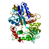 3thiC S: Starting model for refinement C: citing same article ( |
|---|---|
| Similar structure data |


-Links
 PDBj
PDBj


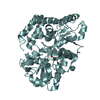
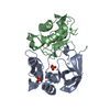
- Assembly
| Deposited unit | 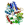
| ||||||||
|---|---|---|---|---|---|---|---|---|---|
| 1 |
| ||||||||
| Unit cell |
| ||||||||
| Components on special symmetry positions |
|
-Components
| #1: Protein | Mass: 40518.801 Da / Num. of mol.: 1 Source method: isolated from a genetically manipulated source Source: (gene. exp.) |
|---|---|
| #2: Chemical | ChemComp-SO4 /   Mass: 96.063 Da / Num. of mol.: 1 / Source method: obtained synthetically / Formula: SO4 Mass: 96.063 Da / Num. of mol.: 1 / Source method: obtained synthetically / Formula: SO4 |
| #3: Chemical | ChemComp-PYD / 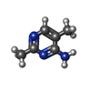  Mass: 123.156 Da / Num. of mol.: 1 / Source method: obtained synthetically / Formula: C6H9N3 Mass: 123.156 Da / Num. of mol.: 1 / Source method: obtained synthetically / Formula: C6H9N3 |
| #4: Water | ChemComp-HOH /  Mass: 18.015 Da / Num. of mol.: 129 / Source method: isolated from a natural source / Formula: H2O Mass: 18.015 Da / Num. of mol.: 129 / Source method: isolated from a natural source / Formula: H2O |
| Has protein modification | Y |
| Nonpolymer details | SULFATE ION ON CRYSTALLOGRAPHIC TWO-FOLD SG OF CYS IS COVALENTLY BOUND TO THE C6 ATOM OF 4-AMINO- ...SULFATE ION ON CRYSTALLOG |
| Sequence details | THE SWISS-PROT SEQUENCE CONTAINS A SIGNAL SEQUENCE THAT GETS PROCESSED AT THREE DIFFERENT SITES. ...THE SWISS-PROT SEQUENCE CONTAINS A SIGNAL SEQUENCE THAT GETS PROCESSED AT THREE DIFFERENT SITES. THE NUMBERING OF RESIDUES IN THIS PDB FILE IS CONSISTENT |
-Experimental details
-Experiment
| Experiment | Method: X-RAY DIFFRACTION / Number of used crystals: 1 |
|---|
- Sample preparation
| Crystal | Density Matthews: 2.29 Å3/Da / Density % sol: 46 % | ||||||||||||||||||||
|---|---|---|---|---|---|---|---|---|---|---|---|---|---|---|---|---|---|---|---|---|---|
| Crystal grow | pH: 4.6 / Details: pH 4.60 | ||||||||||||||||||||
| Crystal grow | *PLUS Method: vapor diffusion, hanging drop | ||||||||||||||||||||
| Components of the solutions | *PLUS
|
-Data collection
| Diffraction | Mean temperature: 100 K |
|---|---|
| Diffraction source | Source: SYNCHROTRON / Site: CHESS  / Beamline: A1 / Wavelength: 0.9104 / Beamline: A1 / Wavelength: 0.9104 |
| Detector | Type: PRINCETON / Detector: CCD |
| Radiation | Protocol: SINGLE WAVELENGTH / Monochromatic (M) / Laue (L): M / Scattering type: x-ray |
| Radiation wavelength | Wavelength: 0.9104 Å / Relative weight: 1 |
| Reflection | Resolution: 2→30 Å / Num. obs: 23969 / % possible obs: 89.2 % / Redundancy: 6.7 % / Rsym value: 0.046 / Net I/σ(I): 31.2 |
| Reflection shell | Resolution: 2→2.07 Å / Redundancy: 1.5 % / Mean I/σ(I) obs: 7.2 / Rsym value: 0.15 / % possible all: 54 |
| Reflection | *PLUS Rmerge(I) obs: 0.046 |
| Reflection shell | *PLUS % possible obs: 54 % / Rmerge(I) obs: 0.15 |
- Processing
| Software |
| ||||||||||||||||||||||||||||||||||||||||||||||||||||||||||||
|---|---|---|---|---|---|---|---|---|---|---|---|---|---|---|---|---|---|---|---|---|---|---|---|---|---|---|---|---|---|---|---|---|---|---|---|---|---|---|---|---|---|---|---|---|---|---|---|---|---|---|---|---|---|---|---|---|---|---|---|---|---|
| Refinement | Method to determine structure: MOLECULAR REPLACEMENT Starting model: RM TEMP STRUCTURE 2THI Resolution: 2→30 Å / Cross valid method: THROUGHOUT / σ(F): 2
| ||||||||||||||||||||||||||||||||||||||||||||||||||||||||||||
| Refinement step | Cycle: LAST / Resolution: 2→30 Å
| ||||||||||||||||||||||||||||||||||||||||||||||||||||||||||||
| Refine LS restraints |
|
