 Movie
Movie Controller

[English] 日本語
 Yorodumi
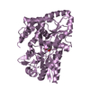
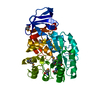
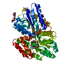
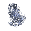
- PDB-2b3b: Thermus thermophilus Glucose/Galactose Binding Protein With Bound... -
+ Open data

- Basic information
| Entry | Database: PDB / ID: 2b3b | ||||||
|---|---|---|---|---|---|---|---|
| Title | Thermus thermophilus Glucose/Galactose Binding Protein With Bound Glucose | ||||||
 Components Components | glucose-binding protein | ||||||
 Keywords Keywords | SUGAR BINDING PROTEIN / protein-carbohydrate complex / glucose / galactose / periplasmic binding protein | ||||||
| Function / homology |  Function and homology information Function and homology information | ||||||
| Biological species |   Thermus thermophilus HB27 (bacteria) Thermus thermophilus HB27 (bacteria) | ||||||
| Method |  X-RAY DIFFRACTION / SYNCHROTRON / SAD / Resolution: 1.95 Å X-RAY DIFFRACTION / SYNCHROTRON / SAD / Resolution: 1.95 Å | ||||||
 Authors Authors | Cuneo, M.J. / Changela, A. / Warren, J.J. / Beese, L.S. / Hellinga, H.W. | ||||||
 Citation Citation | Journal: J.Mol.Biol. / Year: 2006 Title: The crystal structure of a thermophilic glucose binding protein reveals adaptations that interconvert mono and di-saccharide binding sites. Authors: Cuneo, M.J. / Changela, A. / Warren, J.J. / Beese, L.S. / Hellinga, H.W. | ||||||
| History |
| ||||||
| Remark 999 | SEQEUNCE THE AUTHORS STATE THAT THE DISCREPANCIES BETWEEN GENBANK AND AMINO ACID SEQUENCE IN PDB ...SEQEUNCE THE AUTHORS STATE THAT THE DISCREPANCIES BETWEEN GENBANK AND AMINO ACID SEQUENCE IN PDB ARE DUE TO ERRORS IN THE PUBLISHED SEQUENCE OR ERRORS AS A RESULT OF CLONING THE GENE FOR CRYSTALLIZATION. |
- Structure visualization
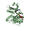
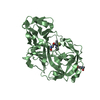
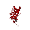
| Structure viewer | Molecule: MolmilJmol/JSmol |
|---|
- Downloads & links
-Download
| PDBx/mmCIF format | 2b3b.cif.gz | 489.8 KB | Display | PDBx/mmCIF format |
|---|---|---|---|---|
| PDB format | pdb2b3b.ent.gz | 401.2 KB | Display | PDB format |
| PDBx/mmJSON format | 2b3b.json.gz | Tree view | PDBx/mmJSON format | |
| Others |  Other downloads Other downloads |
-Validation report
| Arichive directory | https://data.pdbj.org/pub/pdb/validation_reports/b3/2b3bftp://data.pdbj.org/pub/pdb/validation_reports/b3/2b3b | HTTPS FTP |
|---|
-Related structure data


-Links
 PDBj
PDBj


- Assembly
| Deposited unit | 
| ||||||||
|---|---|---|---|---|---|---|---|---|---|
| 1 | 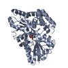
| ||||||||
| 2 | 
| ||||||||
| 3 | 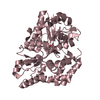
| ||||||||
| 4 | 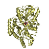
| ||||||||
| 5 | 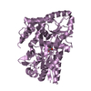
| ||||||||
| 6 | 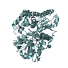
| ||||||||
| Unit cell |
|
-Components
| #1: Protein | Mass: 44091.215 Da / Num. of mol.: 6 Source method: isolated from a genetically manipulated source Source: (gene. exp.) Thermus thermophilus HB27 (bacteria) / Species: Thermus thermophilus / Strain: HB27 / ATCC BAA-163 / DSM 7039 / Gene: ttc0328 / Plasmid: pET21a / Production host: #2: Sugar | ChemComp-GLC /   Type: D-saccharide, alpha linking / Mass: 180.156 Da / Num. of mol.: 4 Type: D-saccharide, alpha linking / Mass: 180.156 Da / Num. of mol.: 4Source method: isolated from a genetically manipulated source Formula: C6H12O6 #3: Sugar |   Type: D-saccharide, beta linking / Mass: 180.156 Da / Num. of mol.: 2 Type: D-saccharide, beta linking / Mass: 180.156 Da / Num. of mol.: 2Source method: isolated from a genetically manipulated source Formula: C6H12O6 #4: Water | ChemComp-HOH / |  Mass: 18.015 Da / Num. of mol.: 1937 / Source method: isolated from a natural source / Formula: H2O Mass: 18.015 Da / Num. of mol.: 1937 / Source method: isolated from a natural source / Formula: H2O |
|---|
-Experimental details
-Experiment
| Experiment | Method: X-RAY DIFFRACTION / Number of used crystals: 1 |
|---|
- Sample preparation
| Crystal | Density Matthews: 2.6 Å3/Da / Density % sol: 52 % |
|---|---|
| Crystal grow | Temperature: 290 K / Method: vapor diffusion, hanging drop / pH: 5.6 Details: 10% (w/v) Isopropanol, 0.1M Sodium Citrate pH5.6, 10% PEG 4000, VAPOR DIFFUSION, HANGING DROP, temperature 290K |
-Data collection
| Diffraction | Mean temperature: 100 K |
|---|---|
| Diffraction source | Source: SYNCHROTRON / Site: APS  / Beamline: 22-ID / Wavelength: 0.97917 Å / Beamline: 22-ID / Wavelength: 0.97917 Å |
| Detector | Type: MARMOSAIC 300 mm CCD / Detector: CCD / Date: Mar 16, 2005 |
| Radiation | Protocol: SINGLE WAVELENGTH / Monochromatic (M) / Laue (L): M / Scattering type: x-ray |
| Radiation wavelength | Wavelength: 0.97917 Å / Relative weight: 1 |
| Reflection | Resolution: 1.95→50 Å / Num. obs: 181610 / % possible obs: 95.6 % / Redundancy: 5.4 % / Rmerge(I) obs: 0.086 / Χ2: 1.767 |
| Reflection shell | Resolution: 1.95→2.02 Å / % possible obs: 82.4 % / Redundancy: 3.8 % / Rmerge(I) obs: 0.455 / Mean I/σ(I) obs: 2.4 / Num. measured obs: 15463 / Χ2: 0.818 / % possible all: 82.4 |
- Processing
| Software |
| ||||||||||||||||||||||||||||||||||||||||||||||||||||||||||||||||||||||||||||||||||||||||||||||||||||||||||||||||||||||||||||||||||
|---|---|---|---|---|---|---|---|---|---|---|---|---|---|---|---|---|---|---|---|---|---|---|---|---|---|---|---|---|---|---|---|---|---|---|---|---|---|---|---|---|---|---|---|---|---|---|---|---|---|---|---|---|---|---|---|---|---|---|---|---|---|---|---|---|---|---|---|---|---|---|---|---|---|---|---|---|---|---|---|---|---|---|---|---|---|---|---|---|---|---|---|---|---|---|---|---|---|---|---|---|---|---|---|---|---|---|---|---|---|---|---|---|---|---|---|---|---|---|---|---|---|---|---|---|---|---|---|---|---|---|---|
| Refinement | Method to determine structure: SAD / Resolution: 1.95→50 Å / Cor.coef. Fo:Fc: 0.961 / Cor.coef. Fo:Fc free: 0.947 / SU B: 3.81 / SU ML: 0.108 / Cross valid method: THROUGHOUT / σ(F): 0 / σ(I): 2.4 / ESU R: 0.175 / ESU R Free: 0.155 / Stereochemistry target values: MAXIMUM LIKELIHOOD / Details: HYDROGENS HAVE BEEN ADDED IN THE RIDING POSITIONS
| ||||||||||||||||||||||||||||||||||||||||||||||||||||||||||||||||||||||||||||||||||||||||||||||||||||||||||||||||||||||||||||||||||
| Solvent computation | Ion probe radii: 0.8 Å / Shrinkage radii: 0.8 Å / VDW probe radii: 1.2 Å / Solvent model: MASK | ||||||||||||||||||||||||||||||||||||||||||||||||||||||||||||||||||||||||||||||||||||||||||||||||||||||||||||||||||||||||||||||||||
| Displacement parameters | Biso mean: 23.086 Å2
| ||||||||||||||||||||||||||||||||||||||||||||||||||||||||||||||||||||||||||||||||||||||||||||||||||||||||||||||||||||||||||||||||||
| Refinement step | Cycle: LAST / Resolution: 1.95→50 Å
| ||||||||||||||||||||||||||||||||||||||||||||||||||||||||||||||||||||||||||||||||||||||||||||||||||||||||||||||||||||||||||||||||||
| Refine LS restraints |
| ||||||||||||||||||||||||||||||||||||||||||||||||||||||||||||||||||||||||||||||||||||||||||||||||||||||||||||||||||||||||||||||||||
| LS refinement shell | Resolution: 1.953→2.004 Å / Total num. of bins used: 20
|
