- PDB-3oo7: Crystal structures and biochemical characterization of the bacter... -
+
Open data
ID or keywords:
Loading...
-
Basic information
Entry
Database: PDB / ID: 3oo7
Title
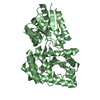
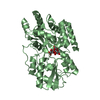
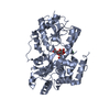
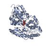
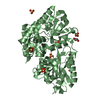
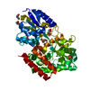
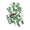
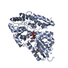
Crystal structures and biochemical characterization of the bacterial solute receptor AcbH reveal an unprecedented exclusive substrate preference for b-D-galactopyranose
Components
ABC transporter binding protein AcbH
Keywords
SUGAR BINDING PROTEIN / class 2 SBP fold / ABC transporter extracellular solute binding protein / D-galactose Binding
Monochromator: Si 111 CHANNEL / Protocol: SINGLE WAVELENGTH / Monochromatic (M) / Laue (L): M / Scattering type: x-ray
Radiation wavelength
Wavelength: 0.91841 Å / Relative weight: 1
Reflection
Number: 192262 / Rmerge(I) obs: 0.158 / D res high: 1.96 Å / Num. obs: 26593 / % possible obs: 100
Diffraction reflection shell
Highest resolution (Å)
Lowest resolution (Å)
Num. obs
% possible obs (%)
ID
Rmerge(I) obs
1.96
2.01
1910
100
1
0.759
2.01
2.07
1898
100
1
0.629
2.07
2.13
1817
100
1
0.556
2.13
2.19
1788
99.9
1
0.452
2.19
2.26
1732
100
1
0.4
2.26
2.34
1676
100
1
0.358
2.34
2.43
1626
100
1
0.317
2.43
2.53
1554
100
1
0.286
2.53
2.64
1525
100
1
0.257
2.64
2.77
1424
100
1
0.203
2.77
2.92
1365
99.9
1
0.181
2.92
3.1
1304
100
1
0.14
3.1
3.31
1246
100
1
0.11
3.31
3.58
1147
100
1
0.083
3.58
3.92
1072
100
1
0.069
3.92
4.38
967
100
1
0.057
4.38
5.06
860
100
1
0.054
5.06
6.2
737
100
1
0.058
6.2
8.77
592
100
1
0.042
Reflection
Resolution: 1.96→34.03 Å / Num. obs: 26593 / % possible obs: 100 % / Observed criterion σ(I): -3 / Redundancy: 7.2 % / Biso Wilson estimate: 23.675 Å2 / Rmerge(I) obs: 0.158 / Net I/σ(I): 14.53
Reflection shell
Diffraction-ID: 1
Resolution (Å)
Highest resolution (Å)
Rmerge F obs
Rmerge(I) obs
Mean I/σ(I) obs
Num. measured obs
Num. possible
Num. unique obs
Rrim(I) all
% possible all
1.96-2.01
0.537
0.759
3.7
13768
1910
1910
0.817
100
2.01-2.07
0.423
0.629
4.5
13795
1898
1898
0.677
100
2.07-2.13
0.351
0.556
5.2
13226
1817
1817
0.598
100
2.13-2.19
0.292
0.452
6.3
13075
1789
1788
0.486
99.9
2.19-2.26
0.252
0.4
7.2
12667
1732
1732
0.43
100
2.26-2.34
0.235
0.358
8
12282
1676
1676
0.385
100
2.34-2.43
0.2
0.317
9.1
11960
1626
1626
0.341
100
2.43-2.53
0.19
0.286
9.9
11354
1554
1554
0.308
100
2.53-2.64
0.167
0.257
10.8
11148
1525
1525
0.276
100
2.64-2.77
0.136
0.203
13
10435
1424
1424
0.219
100
2.77-2.92
0.11
0.181
14.9
9944
1366
1365
0.195
99.9
2.92-3.1
0.081
0.14
18
9480
1304
1304
0.15
100
3.1-3.31
0.063
0.11
22.4
8994
1246
1246
0.118
100
3.31-3.58
0.045
0.083
27.1
8254
1147
1147
0.09
100
3.58-3.92
0.036
0.069
31.5
7681
1072
1072
0.074
100
3.92-4.38
0.029
0.057
35.5
6844
967
967
0.062
100
4.38-5.06
0.03
0.054
35.5
6051
860
860
0.058
100
5.06-6.2
0.034
0.058
31
5148
737
737
0.063
100
6.2-8.77
0.037
0.042
34.1
3996
592
592
0.046
100
8.77
0.018
0.03
42.2
2160
361
353
0.033
97.8
-
Processing
Software
Name
Version
Classification
NB
XSCALE
datascaling
REFMAC
refinement
PDB_EXTRACT
3.1
dataextraction
ADSC
Quantum
datacollection
PHASER
phasing
Refinement
Method to determine structure: MOLECULAR REPLACEMENT Starting model: model from SeMet SAD phasing Resolution: 2.1→33.58 Å / Cor.coef. Fo:Fc: 0.951 / Cor.coef. Fo:Fc free: 0.916 / SU B: 4.91 / SU ML: 0.13 / Cross valid method: THROUGHOUT / σ(F): 0 / ESU R: 0.244 / ESU R Free: 0.191 / Stereochemistry target values: MAXIMUM LIKELIHOOD / Details: HYDROGENS HAVE BEEN ADDED IN THE RIDING POSITIONS
Rfactor
Num. reflection
% reflection
Selection details
Rfree
0.21994
1086
5 %
RANDOM
Rwork
0.16412
-
-
-
obs
0.1669
20648
100 %
-
Solvent computation
Ion probe radii: 0.8 Å / Shrinkage radii: 0.8 Å / VDW probe radii: 1.4 Å / Solvent model: MASK
Displacement parameters
Biso mean: 17.785 Å2
Baniso -1
Baniso -2
Baniso -3
1-
-0.1 Å2
0 Å2
0 Å2
2-
-
0.23 Å2
0 Å2
3-
-
-
-0.13 Å2
Refinement step
Cycle: LAST / Resolution: 2.1→33.58 Å
Protein
Nucleic acid
Ligand
Solvent
Total
Num. atoms
3039
0
56
308
3403
Refine LS restraints
Refine-ID
Type
Dev ideal
Dev ideal target
Number
X-RAY DIFFRACTION
r_bond_refined_d
0.009
0.022
3174
X-RAY DIFFRACTION
r_bond_other_d
X-RAY DIFFRACTION
r_angle_refined_deg
1.248
1.95
4312
X-RAY DIFFRACTION
r_angle_other_deg
X-RAY DIFFRACTION
r_dihedral_angle_1_deg
4.034
5
389
X-RAY DIFFRACTION
r_dihedral_angle_2_deg
32.236
25.347
144
X-RAY DIFFRACTION
r_dihedral_angle_3_deg
13.452
15
496
X-RAY DIFFRACTION
r_dihedral_angle_4_deg
14.472
15
7
X-RAY DIFFRACTION
r_chiral_restr
0.107
0.2
457
X-RAY DIFFRACTION
r_gen_planes_refined
0.014
0.021
2425
X-RAY DIFFRACTION
r_gen_planes_other
X-RAY DIFFRACTION
r_nbd_refined
X-RAY DIFFRACTION
r_nbd_other
X-RAY DIFFRACTION
r_nbtor_refined
X-RAY DIFFRACTION
r_nbtor_other
X-RAY DIFFRACTION
r_xyhbond_nbd_refined
X-RAY DIFFRACTION
r_xyhbond_nbd_other
X-RAY DIFFRACTION
r_metal_ion_refined
X-RAY DIFFRACTION
r_metal_ion_other
X-RAY DIFFRACTION
r_symmetry_vdw_refined
X-RAY DIFFRACTION
r_symmetry_vdw_other
X-RAY DIFFRACTION
r_symmetry_hbond_refined
X-RAY DIFFRACTION
r_symmetry_hbond_other
X-RAY DIFFRACTION
r_symmetry_metal_ion_refined
X-RAY DIFFRACTION
r_symmetry_metal_ion_other
X-RAY DIFFRACTION
r_mcbond_it
1.158
1.5
1941
X-RAY DIFFRACTION
r_mcbond_other
X-RAY DIFFRACTION
r_mcangle_it
1.844
2
3120
X-RAY DIFFRACTION
r_scbond_it
3.372
3
1233
X-RAY DIFFRACTION
r_scangle_it
4.945
4.5
1192
X-RAY DIFFRACTION
r_rigid_bond_restr
X-RAY DIFFRACTION
r_sphericity_free
X-RAY DIFFRACTION
r_sphericity_bonded
LS refinement shell
Resolution: 2.1→2.154 Å / Total num. of bins used: 20
Rfactor
Num. reflection
% reflection
Rfree
0.256
78
-
Rwork
0.203
1478
-
obs
-
-
100 %
+
About Yorodumi
-
News
-
Feb 9, 2022. New format data for meta-information of EMDB entries
New format data for meta-information of EMDB entries
Version 3 of the EMDB header file is now the official format.
The previous official version 1.9 will be removed from the archive.
In the structure databanks used in Yorodumi, some data are registered as the other names, "COVID-19 virus" and "2019-nCoV". Here are the details of the virus and the list of structure data.
Jan 31, 2019. EMDB accession codes are about to change! (news from PDBe EMDB page)
EMDB accession codes are about to change! (news from PDBe EMDB page)
The allocation of 4 digits for EMDB accession codes will soon come to an end. Whilst these codes will remain in use, new EMDB accession codes will include an additional digit and will expand incrementally as the available range of codes is exhausted. The current 4-digit format prefixed with “EMD-” (i.e. EMD-XXXX) will advance to a 5-digit format (i.e. EMD-XXXXX), and so on. It is currently estimated that the 4-digit codes will be depleted around Spring 2019, at which point the 5-digit format will come into force.
The EM Navigator/Yorodumi systems omit the EMD- prefix.
Related info.:Q: What is EMD? / ID/Accession-code notation in Yorodumi/EM Navigator
Yorodumi is a browser for structure data from EMDB, PDB, SASBDB, etc.
This page is also the successor to EM Navigator detail page, and also detail information page/front-end page for Omokage search.
The word "yorodu" (or yorozu) is an old Japanese word meaning "ten thousand". "mi" (miru) is to see.
Related info.:EMDB / PDB / SASBDB / Comparison of 3 databanks / Yorodumi Search / Aug 31, 2016. New EM Navigator & Yorodumi / Yorodumi Papers / Jmol/JSmol / Function and homology information / Changes in new EM Navigator and Yorodumi
 Movie
Movie Controller

 Yorodumi
 Open data
 Basic information
 Components
 Keywords
 Function and homology information
 Actinoplanes (bacteria)
 X-RAY DIFFRACTION /
  Authors
 Citation
 Structure visualization
 Downloads & links
 Other downloads


 PDBj
PDBj


 Assembly

 Mass: 96.063 Da / Num. of mol.: 4 / Source method: obtained synthetically / Formula: SO4
 Mass: 92.094 Da / Num. of mol.: 6 / Source method: obtained synthetically / Formula: C3H8O3
 Mass: 18.015 Da / Num. of mol.: 308 / Source method: isolated from a natural source / Formula: H2O
 Sample preparation
 / Beamline: 14.2 / Wavelength: 0.91841 Å
 Processing