 Movie
Movie Controller


+ Open data

- Basic information
| Entry | Database: PDB / ID: 6pql | |||||||||
|---|---|---|---|---|---|---|---|---|---|---|
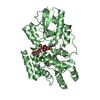
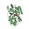
| Title | SBP RafE in complex with raffinose | |||||||||
 Components Components | ABC transporter sugar-binding protein | |||||||||
 Keywords Keywords | SUGAR BINDING PROTEIN / Cluster D-I solute binding protein / complex / SBP | |||||||||
| Function / homology |  Function and homology information Function and homology information | |||||||||
| Biological species |  Streptococcus pneumoniae TIGR4 (bacteria) Streptococcus pneumoniae TIGR4 (bacteria) | |||||||||
| Method |  X-RAY DIFFRACTION / MOLECULAR REPLACEMENT / molecular replacement / Resolution: 2.65 Å X-RAY DIFFRACTION / MOLECULAR REPLACEMENT / molecular replacement / Resolution: 2.65 Å | |||||||||
 Authors Authors | Meier, E.P.W. / Boraston, A.B. | |||||||||
| Funding support |  Canada, 1items Canada, 1items
| |||||||||
 Citation Citation | Journal: J.Biol.Chem. / Year: 2019 Title: Molecular analysis of an enigmaticStreptococcus pneumoniaevirulence factor: The raffinose-family oligosaccharide utilization system. Authors: Hobbs, J.K. / Meier, E.P.W. / Pluvinage, B. / Mey, M.A. / Boraston, A.B. | |||||||||
| History |
|
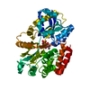
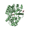
- Structure visualization
| Structure viewer | Molecule: MolmilJmol/JSmol |
|---|
- Downloads & links
-Download
| PDBx/mmCIF format | 6pql.cif.gz | 304.4 KB | Display | PDBx/mmCIF format |
|---|---|---|---|---|
| PDB format | pdb6pql.ent.gz | 243.9 KB | Display | PDB format |
| PDBx/mmJSON format | 6pql.json.gz | Tree view | PDBx/mmJSON format | |
| Others |  Other downloads Other downloads |
-Validation report
| Arichive directory | https://data.pdbj.org/pub/pdb/validation_reports/pq/6pqlftp://data.pdbj.org/pub/pdb/validation_reports/pq/6pql | HTTPS FTP |
|---|
-Related structure data
| Related structure data | 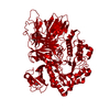 6phuC 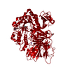 6phvC 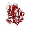 6phwC 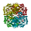 6phxC 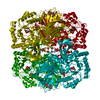 6phyC 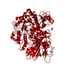 6pi0C 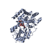 6preC 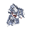 6prgSC S: Starting model for refinement C: citing same article ( |
|---|---|
| Similar structure data |


-Links
 PDBj
PDBj


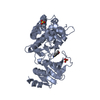
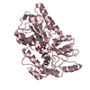
- Assembly
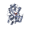
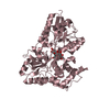
| Deposited unit | 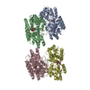
| ||||||||
|---|---|---|---|---|---|---|---|---|---|
| 1 | 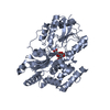
| ||||||||
| 2 | 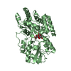
| ||||||||
| 3 | 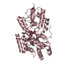
| ||||||||
| 4 | 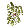
| ||||||||
| Unit cell |
|
-Components
| #1: Protein | Mass: 46440.012 Da / Num. of mol.: 4 Source method: isolated from a genetically manipulated source Source: (gene. exp.) Streptococcus pneumoniae TIGR4 (bacteria)Gene: SP_1897 / Plasmid: pET28a / Production host: #2: Polysaccharide | beta-D-fructofuranose-(2-1)-[alpha-D-galactopyranose-(1-6)]alpha-D-glucopyranose Source method: isolated from a genetically manipulated source #3: Water | ChemComp-HOH / |  Mass: 18.015 Da / Num. of mol.: 239 / Source method: isolated from a natural source / Formula: H2O Mass: 18.015 Da / Num. of mol.: 239 / Source method: isolated from a natural source / Formula: H2OHas ligand of interest | Y | |
|---|
-Experimental details
-Experiment
| Experiment | Method: X-RAY DIFFRACTION / Number of used crystals: 1 |
|---|
- Sample preparation
| Crystal | Density Matthews: 2.27 Å3/Da / Density % sol: 45.9 % |
|---|---|
| Crystal grow | Temperature: 291 K / Method: vapor diffusion, sitting drop / pH: 8 / Details: 0.2M CsCl, 0.1M Tris, 20% w/v PEG 3350 |
-Data collection
| Diffraction | Mean temperature: 100 K / Serial crystal experiment: N |
|---|---|
| Diffraction source | Source: ROTATING ANODE / Type: RIGAKU MICROMAX-002 / Wavelength: 1.54187 Å |
| Detector | Type: DECTRIS PILATUS 200K / Detector: PIXEL / Date: Jan 21, 2017 |
| Radiation | Protocol: SINGLE WAVELENGTH / Monochromatic (M) / Laue (L): M / Scattering type: x-ray |
| Radiation wavelength | Wavelength: 1.54187 Å / Relative weight: 1 |
| Reflection | Resolution: 2.65→30 Å / Num. obs: 47919 / % possible obs: 100 % / Redundancy: 4 % / CC1/2: 0.987 / Rmerge(I) obs: 0.142 / Rpim(I) all: 0.083 / Net I/σ(I): 11 |
| Reflection shell | Resolution: 2.65→2.7 Å / Rmerge(I) obs: 0.565 / Num. unique obs: 4257 / CC1/2: 0.729 / Rpim(I) all: 0.369 |
-Phasing
| Phasing | Method: molecular replacement |
|---|
- Processing
| Software |
| |||||||||||||||||||||||||||||||||||||||||||||
|---|---|---|---|---|---|---|---|---|---|---|---|---|---|---|---|---|---|---|---|---|---|---|---|---|---|---|---|---|---|---|---|---|---|---|---|---|---|---|---|---|---|---|---|---|---|---|
| Refinement | Method to determine structure: MOLECULAR REPLACEMENT Starting model: 6PRG Resolution: 2.65→29.66 Å / Cor.coef. Fo:Fc: 0.895 / Cor.coef. Fo:Fc free: 0.847 / SU B: 16.079 / SU ML: 0.331 / Cross valid method: THROUGHOUT / σ(F): 0 / ESU R Free: 0.414 / Details: U VALUES : REFINED INDIVIDUALLY
| |||||||||||||||||||||||||||||||||||||||||||||
| Solvent computation | Ion probe radii: 0.8 Å / Shrinkage radii: 0.8 Å / VDW probe radii: 1.2 Å | |||||||||||||||||||||||||||||||||||||||||||||
| Displacement parameters | Biso max: 84.55 Å2 / Biso mean: 39.709 Å2 / Biso min: 6.7 Å2
| |||||||||||||||||||||||||||||||||||||||||||||
| Refinement step | Cycle: final / Resolution: 2.65→29.66 Å
| |||||||||||||||||||||||||||||||||||||||||||||
| Refine LS restraints |
| |||||||||||||||||||||||||||||||||||||||||||||
| LS refinement shell | Resolution: 2.65→2.719 Å / Rfactor Rfree error: 0 / Total num. of bins used: 20
|
