 Movie
Movie Controller

[English] 日本語
 Yorodumi
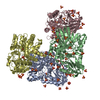
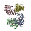
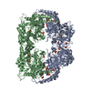
- PDB-3on5: Crystal structure of a xanthine dehydrogenase (BH1974) from Bacil... -
+ Open data

- Basic information
| Entry | Database: PDB / ID: 3on5 | ||||||
|---|---|---|---|---|---|---|---|
| Title | Crystal structure of a xanthine dehydrogenase (BH1974) from Bacillus halodurans at 2.80 A resolution | ||||||
 Components Components | BH1974 protein | ||||||
 Keywords Keywords | OXIDOREDUCTASE / STRUCTURAL GENOMICS / JOINT CENTER FOR STRUCTURAL GENOMICS / JCSG / PROTEIN STRUCTURE INITIATIVE / PSI-BIOLOGY | ||||||
| Function / homology |  Function and homology information Function and homology information: / BH1974-like, central domain / XdhC- CoxI / XdhC Rossmann domain / : / XdhC and CoxI family / XdhC Rossmann domain / NAD(P)-binding Rossmann-like Domain / Rossmann fold / 3-Layer(aba) Sandwich / Alpha Beta Similarity search - Domain/homology | ||||||
| Biological species |  Bacillus halodurans (bacteria) Bacillus halodurans (bacteria) | ||||||
| Method |  X-RAY DIFFRACTION / SYNCHROTRON / MAD, MOLECULAR REPLACEMENT / MAD, molecular replacement / Resolution: 2.8 Å X-RAY DIFFRACTION / SYNCHROTRON / MAD, MOLECULAR REPLACEMENT / MAD, molecular replacement / Resolution: 2.8 Å | ||||||
 Authors Authors | Joint Center for Structural Genomics (JCSG) | ||||||
 Citation Citation | Journal: To be published Title: Crystal structure of a xanthine dehydrogenase (BH1974) from Bacillus halodurans at 2.80 A resolution Authors: Joint Center for Structural Genomics (JCSG) | ||||||
| History |
|
- Structure visualization
| Structure viewer | Molecule: MolmilJmol/JSmol |
|---|
- Downloads & links
-Download
| PDBx/mmCIF format | 3on5.cif.gz | 275.5 KB | Display | PDBx/mmCIF format |
|---|---|---|---|---|
| PDB format | pdb3on5.ent.gz | 225.4 KB | Display | PDB format |
| PDBx/mmJSON format | 3on5.json.gz | Tree view | PDBx/mmJSON format | |
| Others |  Other downloads Other downloads |
-Validation report
| Arichive directory | https://data.pdbj.org/pub/pdb/validation_reports/on/3on5ftp://data.pdbj.org/pub/pdb/validation_reports/on/3on5 | HTTPS FTP |
|---|
-Related structure data
| Related structure data |  2we8S S: Starting model for refinement |
|---|---|
| Similar structure data | |
| Other databases |


-Links
 PDBj
PDBj- Assembly
| Deposited unit | 
| ||||||||
|---|---|---|---|---|---|---|---|---|---|
| 1 | 
| ||||||||
| 2 |
| ||||||||
| Unit cell |
|
-Components
| #1: Protein | Mass: 41786.445 Da / Num. of mol.: 2 Source method: isolated from a genetically manipulated source Source: (gene. exp.) Bacillus halodurans (bacteria) / Gene: 10174592, BH1974 / Plasmid: SpeedET / Production host: #2: Chemical |   Mass: 35.453 Da / Num. of mol.: 2 / Source method: obtained synthetically / Formula: Cl Mass: 35.453 Da / Num. of mol.: 2 / Source method: obtained synthetically / Formula: Cl#3: Water | ChemComp-HOH / |  Mass: 18.015 Da / Num. of mol.: 51 / Source method: isolated from a natural source / Formula: H2O Mass: 18.015 Da / Num. of mol.: 51 / Source method: isolated from a natural source / Formula: H2OHas protein modification | Y | Sequence details | THE CONSTRUCT WAS EXPRESSED WITH A PURIFICATI | |
|---|
-Experimental details
-Experiment
| Experiment | Method: X-RAY DIFFRACTION / Number of used crystals: 1 |
|---|
- Sample preparation
| Crystal | Density Matthews: 3.26 Å3/Da / Density % sol: 62.29 % Description: INITIAL PHASING WAS PERFORMED USING A ROSETTA-BASED APPROACH. PHASER WAS USED TO FIND AN ENSEMBLE OF CANDIDATE MOLECULAR REPLACEMENT SOLUTIONS USING COMPARATIVE MODELS BUILT BY ROSETTA ...Description: INITIAL PHASING WAS PERFORMED USING A ROSETTA-BASED APPROACH. PHASER WAS USED TO FIND AN ENSEMBLE OF CANDIDATE MOLECULAR REPLACEMENT SOLUTIONS USING COMPARATIVE MODELS BUILT BY ROSETTA BASED ON PDB ID 2WE8. ROSETTA WAS THEN USED TO REFINE THE CANDIDATE SOLUTION INTO DENSITY. THE SE SITES FROM THE MOLECULAR REPLACEMENT SOLUTION WERE USED AS AN INITIAL HEAVY ATOM MODEL FOR TWO-WAVELENGTH MAD PHASE REFINEMENT USING SHARP. |
|---|---|
| Crystal grow | Temperature: 293 K / Method: vapor diffusion, sitting drop / pH: 5 Details: 5.0% PEG-6000, 1.1M LiCl, 0.1M Citrate pH 5.0, NANODROP, VAPOR DIFFUSION, SITTING DROP, temperature 293K |
-Data collection
| Diffraction | Mean temperature: 100 K | |||||||||||||||||||||||||||||||||||||||||||||||||||||||||||||||||||||||||||||
|---|---|---|---|---|---|---|---|---|---|---|---|---|---|---|---|---|---|---|---|---|---|---|---|---|---|---|---|---|---|---|---|---|---|---|---|---|---|---|---|---|---|---|---|---|---|---|---|---|---|---|---|---|---|---|---|---|---|---|---|---|---|---|---|---|---|---|---|---|---|---|---|---|---|---|---|---|---|---|
| Diffraction source | Source: SYNCHROTRON / Site: SSRL  / Beamline: BL11-1 / Wavelength: 0.978954,0.918370 / Beamline: BL11-1 / Wavelength: 0.978954,0.918370 | |||||||||||||||||||||||||||||||||||||||||||||||||||||||||||||||||||||||||||||
| Detector | Type: ADSC QUANTUM 315 / Detector: CCD / Date: Jun 4, 2006 / Details: Flat mirror (vertical focusing) | |||||||||||||||||||||||||||||||||||||||||||||||||||||||||||||||||||||||||||||
| Radiation | Monochromator: Single crystal Si(111) bent monochromator (horizontal focusing) Protocol: MAD / Monochromatic (M) / Laue (L): M / Scattering type: x-ray | |||||||||||||||||||||||||||||||||||||||||||||||||||||||||||||||||||||||||||||
| Radiation wavelength |
| |||||||||||||||||||||||||||||||||||||||||||||||||||||||||||||||||||||||||||||
| Reflection | Resolution: 2.8→94.244 Å / Num. all: 28218 / Num. obs: 28218 / % possible obs: 99.9 % / Redundancy: 6.5 % / Biso Wilson estimate: 72.405 Å2 / Rsym value: 0.13 / Net I/σ(I): 12.6 | |||||||||||||||||||||||||||||||||||||||||||||||||||||||||||||||||||||||||||||
| Reflection shell | Rmerge(I) obs: 0.011 / Diffraction-ID: 1
|
-Phasing
| Phasing | Method: MAD, molecular replacement |
|---|
- Processing
| Software |
| ||||||||||||||||||||||||||||||||||||||||||||||||||||||||||||||||||||||||||||||||||||||||||||||||||||||||||||
|---|---|---|---|---|---|---|---|---|---|---|---|---|---|---|---|---|---|---|---|---|---|---|---|---|---|---|---|---|---|---|---|---|---|---|---|---|---|---|---|---|---|---|---|---|---|---|---|---|---|---|---|---|---|---|---|---|---|---|---|---|---|---|---|---|---|---|---|---|---|---|---|---|---|---|---|---|---|---|---|---|---|---|---|---|---|---|---|---|---|---|---|---|---|---|---|---|---|---|---|---|---|---|---|---|---|---|---|---|---|
| Refinement | Method to determine structure: MAD, MOLECULAR REPLACEMENT Starting model: 2WE8 Resolution: 2.8→94.244 Å / Cor.coef. Fo:Fc: 0.9361 / Cor.coef. Fo:Fc free: 0.9256 / Occupancy max: 1 / Occupancy min: 0.5 / Cross valid method: THROUGHOUT / σ(F): 0 Details: 1. A MET-INHIBITION PROTOCOL WAS USED FOR SELENOMETHIONINE INCORPORATION DURING PROTEIN EXPRESSION. THE OCCUPANCY OF THE SE ATOMS IN THE MSE RESIDUES WAS REDUCED TO 0.75 FOR THE REDUCED ...Details: 1. A MET-INHIBITION PROTOCOL WAS USED FOR SELENOMETHIONINE INCORPORATION DURING PROTEIN EXPRESSION. THE OCCUPANCY OF THE SE ATOMS IN THE MSE RESIDUES WAS REDUCED TO 0.75 FOR THE REDUCED SCATTERING POWER DUE TO PARTIAL S-MET INCORPORATION. 2. ATOM RECORD CONTAINS SUM OF TLS AND RESIDUAL B FACTORS. ANISOU RECORD CONTAINS SUM OF TLS AND RESIDUAL U FACTORS. 3. ELECTRON DENSITIES CORRESPONDING TO RESIDUES 43-50 AND RESIDUES 84-94 ON THE B SUBUNIT WERE DISORDERED AND THESE REGIONS COULD NOT BE RELIABLY MODELED. 4. UN-EXPLAINED ELECTRON DENSITY NEAR THE SIDECHAIN OF CYS A92 WAS NOT MODELED. 5. THE REFINEMENT WAS RESTRAINED AGAINST THE TWO WAVELENGTH MAD PHASES. 6. NCS RESTRAINTS WERE APPLIED USING BUSTER'S LSSR RESTRAINT REPRESENTATION (-AUTONCS).
| ||||||||||||||||||||||||||||||||||||||||||||||||||||||||||||||||||||||||||||||||||||||||||||||||||||||||||||
| Displacement parameters | Biso max: 171.31 Å2 / Biso mean: 72.5282 Å2 / Biso min: 20.91 Å2
| ||||||||||||||||||||||||||||||||||||||||||||||||||||||||||||||||||||||||||||||||||||||||||||||||||||||||||||
| Refinement step | Cycle: LAST / Resolution: 2.8→94.244 Å
| ||||||||||||||||||||||||||||||||||||||||||||||||||||||||||||||||||||||||||||||||||||||||||||||||||||||||||||
| Refine LS restraints |
| ||||||||||||||||||||||||||||||||||||||||||||||||||||||||||||||||||||||||||||||||||||||||||||||||||||||||||||
| LS refinement shell | Resolution: 2.8→2.91 Å / Total num. of bins used: 14
| ||||||||||||||||||||||||||||||||||||||||||||||||||||||||||||||||||||||||||||||||||||||||||||||||||||||||||||
| Refinement TLS params. | Method: refined / Refine-ID: X-RAY DIFFRACTION
| ||||||||||||||||||||||||||||||||||||||||||||||||||||||||||||||||||||||||||||||||||||||||||||||||||||||||||||
| Refinement TLS group |
|
