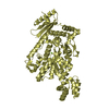
 Movie
Movie Controller

[English] 日本語
 Yorodumi
- PDB-3olm: Structure and Function of a Ubiquitin Binding Site within the Cat... -
+ Open data

- Basic information
| Entry | Database: PDB / ID: 3olm | ||||||
|---|---|---|---|---|---|---|---|
| Title | Structure and Function of a Ubiquitin Binding Site within the Catalytic Domain of a HECT Ubiquitin Ligase | ||||||
 Components Components |
| ||||||
 Keywords Keywords | LIGASE / ubiquitin E3 ligase | ||||||
| Function / homology |  Function and homology information Function and homology informationregulation of dolichyl monophosphate biosynthetic process / regulation of ribosomal large subunit export from nucleus / regulation of tRNA processing / regulation of tRNA export from nucleus / regulation of ubiquinone biosynthetic process / regulation of ergosterol biosynthetic process / positive regulation of ubiquitin-dependent endocytosis / : / RAS processing / Regulation of PTEN localization ...regulation of dolichyl monophosphate biosynthetic process / regulation of ribosomal large subunit export from nucleus / regulation of tRNA processing / regulation of tRNA export from nucleus / regulation of ubiquinone biosynthetic process / regulation of ergosterol biosynthetic process / positive regulation of ubiquitin-dependent endocytosis / : / RAS processing / Regulation of PTEN localization / : / RHOU GTPase cycle / UCH proteinases / PINK1-PRKN Mediated Mitophagy / : / Aggrephagy / Pexophagy / RHOQ GTPase cycle / : / : / cellular response to L-arginine / Peroxisomal protein import / free ubiquitin chain polymerization / : / Endosomal Sorting Complex Required For Transport (ESCRT) / mitochondria-associated ubiquitin-dependent protein catabolic process / regulation of mRNA export from nucleus / late endosome to vacuole transport via multivesicular body sorting pathway / : / actin cortical patch / cellular bud tip / protein transport to vacuole involved in ubiquitin-dependent protein catabolic process via the multivesicular body sorting pathway / regulation of rRNA processing / ribophagy / Translesion synthesis by REV1 / : / : / : / positive regulation of fatty acid biosynthetic process / regulation of nitrogen utilization / : / ubiquitin-dependent protein catabolic process via the multivesicular body sorting pathway / HECT-type E3 ubiquitin transferase / : / Recruitment and ATM-mediated phosphorylation of repair and signaling proteins at DNA double strand breaks / nonfunctional rRNA decay / ubiquitin-dependent endocytosis / Regulation of PTEN stability and activity / poly(A)+ mRNA export from nucleus / ubiquitin-ubiquitin ligase activity / CDK-mediated phosphorylation and removal of Cdc6 / FBXL7 down-regulates AURKA during mitotic entry and in early mitosis / KEAP1-NFE2L2 pathway / Neddylation / cellular response to stress / Ubiquitin-Mediated Degradation of Phosphorylated Cdc25A / Orc1 removal from chromatin / MAPK6/MAPK4 signaling / Formation of TC-NER Pre-Incision Complex / SRP-dependent cotranslational protein targeting to membrane / GTP hydrolysis and joining of the 60S ribosomal subunit / Formation of a pool of free 40S subunits / Nonsense Mediated Decay (NMD) independent of the Exon Junction Complex (EJC) / Nonsense Mediated Decay (NMD) enhanced by the Exon Junction Complex (EJC) / Antigen processing: Ubiquitination & Proteasome degradation / protein quality control for misfolded or incompletely synthesized proteins / L13a-mediated translational silencing of Ceruloplasmin expression / Gap-filling DNA repair synthesis and ligation in TC-NER / Dual incision in TC-NER / positive regulation of endocytosis / Ub-specific processing proteases / protein K63-linked ubiquitination / ubiquitin ligase complex / phosphatidylinositol binding / cytosolic ribosome / regulation of actin cytoskeleton organization / ubiquitin binding / mitochondrion organization / cellular response to amino acid stimulus / positive regulation of receptor-mediated endocytosis / modification-dependent protein catabolic process / protein tag activity / ubiquitin-protein transferase activity / ubiquitin protein ligase activity / peroxisome / regulation of protein localization / positive regulation of proteasomal ubiquitin-dependent protein catabolic process / cellular response to heat / chromatin organization / ubiquitin-dependent protein catabolic process / proteasome-mediated ubiquitin-dependent protein catabolic process / endosome membrane / protein ubiquitination / ubiquitin protein ligase binding / Golgi apparatus / positive regulation of transcription by RNA polymerase II / mitochondrion / nucleus / plasma membrane / cytoplasm Similarity search - Function | ||||||
| Biological species |  | ||||||
| Method |  X-RAY DIFFRACTION / SYNCHROTRON / MOLECULAR REPLACEMENT / Resolution: 2.495 Å X-RAY DIFFRACTION / SYNCHROTRON / MOLECULAR REPLACEMENT / Resolution: 2.495 Å | ||||||
 Authors Authors | Kim, H.C. / Steffen, A. / Chen, J. / Huibregtse, J.M. | ||||||
 Citation Citation | Journal: Embo Rep. / Year: 2011 Title: Structure and function of a HECT domain ubiquitin-binding site. Authors: Kim, H.C. / Steffen, A.M. / Oldham, M.L. / Chen, J. / Huibregtse, J.M. | ||||||
| History |
|
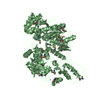
- Structure visualization
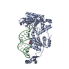
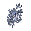
| Structure viewer | Molecule: MolmilJmol/JSmol |
|---|
- Downloads & links
-Download
| PDBx/mmCIF format | 3olm.cif.gz | 211.4 KB | Display | PDBx/mmCIF format |
|---|---|---|---|---|
| PDB format | pdb3olm.ent.gz | 169 KB | Display | PDB format |
| PDBx/mmJSON format | 3olm.json.gz | Tree view | PDBx/mmJSON format | |
| Others |  Other downloads Other downloads |
-Validation report
| Arichive directory | https://data.pdbj.org/pub/pdb/validation_reports/ol/3olmftp://data.pdbj.org/pub/pdb/validation_reports/ol/3olm | HTTPS FTP |
|---|
-Related structure data
| Related structure data |  1nd7S S: Starting model for refinement |
|---|---|
| Similar structure data |


-Links
 PDBj
PDBj


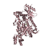
- Assembly
| Deposited unit | 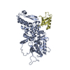
| ||||||||
|---|---|---|---|---|---|---|---|---|---|
| 1 |
| ||||||||
| Unit cell |
|
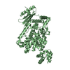
-Components
| #1: Protein | Mass: 50366.660 Da / Num. of mol.: 1 / Fragment: WW3 and HECT domain Source method: isolated from a genetically manipulated source Source: (gene. exp.) Gene: RSP5, MDP1, NPI1, YER125W, SYGP-ORF41 / Production host:  References: UniProt: P39940, Ligases; Forming carbon-nitrogen bonds; Acid-amino-acid ligases (peptide synthases) |
|---|---|
| #2: Protein | Mass: 8841.026 Da / Num. of mol.: 1 Source method: isolated from a genetically manipulated source Source: (gene. exp.) Gene: UBI4, SCD2, YLL039C / Production host: |
| #3: Water | ChemComp-HOH /  Mass: 18.015 Da / Num. of mol.: 51 / Source method: isolated from a natural source / Formula: H2O Mass: 18.015 Da / Num. of mol.: 51 / Source method: isolated from a natural source / Formula: H2O |
-Experimental details
-Experiment
| Experiment | Method: X-RAY DIFFRACTION / Number of used crystals: 1 |
|---|
- Sample preparation
| Crystal | Density Matthews: 2.44 Å3/Da / Density % sol: 49.67 % |
|---|---|
| Crystal grow | Temperature: 293 K / Method: vapor diffusion, sitting drop / pH: 9 Details: 13.5% PEG 4000, 0.2M magnesium chloride, 0.1M Tris-HCl pH 9.0, VAPOR DIFFUSION, SITTING DROP, temperature 293K |
-Data collection
| Diffraction | Mean temperature: 100 K |
|---|---|
| Diffraction source | Source: SYNCHROTRON / Site: APS  / Beamline: 23-ID-D / Wavelength: 1.03326 Å / Beamline: 23-ID-D / Wavelength: 1.03326 Å |
| Detector | Type: MARMOSAIC 300 mm CCD / Detector: CCD / Date: Oct 28, 2009 |
| Radiation | Monochromator: Double crystal cryo-cooled / Protocol: SINGLE WAVELENGTH / Monochromatic (M) / Laue (L): M / Scattering type: x-ray |
| Radiation wavelength | Wavelength: 1.03326 Å / Relative weight: 1 |
| Reflection | Resolution: 2.5→50 Å / Num. obs: 14422 / % possible obs: 75.2 % / Observed criterion σ(F): 0 / Observed criterion σ(I): 0 / Biso Wilson estimate: 38.76 Å2 |
| Reflection shell | Resolution: 2.5→2.59 Å / % possible all: 33.8 |
- Processing
| Software |
| |||||||||||||||||||||||||||||||||||||||||||||||||||||||||||||||||||||||||||||||||||||||||||||||||||||||||||||||||||||||||||||||||||||||||||||||||||||||||||||||||||||||||||||||
|---|---|---|---|---|---|---|---|---|---|---|---|---|---|---|---|---|---|---|---|---|---|---|---|---|---|---|---|---|---|---|---|---|---|---|---|---|---|---|---|---|---|---|---|---|---|---|---|---|---|---|---|---|---|---|---|---|---|---|---|---|---|---|---|---|---|---|---|---|---|---|---|---|---|---|---|---|---|---|---|---|---|---|---|---|---|---|---|---|---|---|---|---|---|---|---|---|---|---|---|---|---|---|---|---|---|---|---|---|---|---|---|---|---|---|---|---|---|---|---|---|---|---|---|---|---|---|---|---|---|---|---|---|---|---|---|---|---|---|---|---|---|---|---|---|---|---|---|---|---|---|---|---|---|---|---|---|---|---|---|---|---|---|---|---|---|---|---|---|---|---|---|---|---|---|---|---|
| Refinement | Method to determine structure: MOLECULAR REPLACEMENT Starting model: 1ND7 Resolution: 2.495→42.085 Å / SU ML: 0.38 / σ(F): 1.34 / Phase error: 31.22 / Stereochemistry target values: ML
| |||||||||||||||||||||||||||||||||||||||||||||||||||||||||||||||||||||||||||||||||||||||||||||||||||||||||||||||||||||||||||||||||||||||||||||||||||||||||||||||||||||||||||||||
| Solvent computation | Shrinkage radii: 0.9 Å / VDW probe radii: 1.11 Å / Solvent model: FLAT BULK SOLVENT MODEL / Bsol: 33.734 Å2 / ksol: 0.321 e/Å3 | |||||||||||||||||||||||||||||||||||||||||||||||||||||||||||||||||||||||||||||||||||||||||||||||||||||||||||||||||||||||||||||||||||||||||||||||||||||||||||||||||||||||||||||||
| Displacement parameters |
| |||||||||||||||||||||||||||||||||||||||||||||||||||||||||||||||||||||||||||||||||||||||||||||||||||||||||||||||||||||||||||||||||||||||||||||||||||||||||||||||||||||||||||||||
| Refinement step | Cycle: LAST / Resolution: 2.495→42.085 Å
| |||||||||||||||||||||||||||||||||||||||||||||||||||||||||||||||||||||||||||||||||||||||||||||||||||||||||||||||||||||||||||||||||||||||||||||||||||||||||||||||||||||||||||||||
| Refine LS restraints |
| |||||||||||||||||||||||||||||||||||||||||||||||||||||||||||||||||||||||||||||||||||||||||||||||||||||||||||||||||||||||||||||||||||||||||||||||||||||||||||||||||||||||||||||||
| LS refinement shell |
| |||||||||||||||||||||||||||||||||||||||||||||||||||||||||||||||||||||||||||||||||||||||||||||||||||||||||||||||||||||||||||||||||||||||||||||||||||||||||||||||||||||||||||||||
| Refinement TLS params. | Method: refined / Refine-ID: X-RAY DIFFRACTION
| |||||||||||||||||||||||||||||||||||||||||||||||||||||||||||||||||||||||||||||||||||||||||||||||||||||||||||||||||||||||||||||||||||||||||||||||||||||||||||||||||||||||||||||||
| Refinement TLS group |
|
