 Movie
Movie Controller

[English] 日本語
 Yorodumi
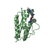
- PDB-3oea: Crystal structure of the Q121E mutants of C.polysaccharolyticus C... -
+ Open data

- Basic information
| Entry | Database: PDB / ID: 3oea | |||||||||
|---|---|---|---|---|---|---|---|---|---|---|
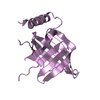
| Title | Crystal structure of the Q121E mutants of C.polysaccharolyticus CBM16-1 bound to cellopentaose | |||||||||
 Components Components | S-layer associated multidomain endoglucanase | |||||||||
 Keywords Keywords | HYDROLASE / Carbohydrate Binding Domain / Cellopentaose | |||||||||
| Function / homology |  Function and homology information Function and homology informationpolysaccharide catabolic process / hydrolase activity, hydrolyzing O-glycosyl compounds / metal ion binding Similarity search - Function | |||||||||
| Biological species |   Caldanaerobius polysaccharolyticus (bacteria) Caldanaerobius polysaccharolyticus (bacteria) | |||||||||
| Method |  X-RAY DIFFRACTION / SYNCHROTRON / MOLECULAR REPLACEMENT / Resolution: 1.35 Å X-RAY DIFFRACTION / SYNCHROTRON / MOLECULAR REPLACEMENT / Resolution: 1.35 Å | |||||||||
 Authors Authors | Agarwal, V. / Nair, S.K. | |||||||||
 Citation Citation | Journal: J.Biol.Chem. / Year: 2010 Title: Mutational insights into the roles of amino acid residues in ligand binding for two closely related family 16 carbohydrate binding modules. Authors: Su, X. / Agarwal, V. / Dodd, D. / Bae, B. / Mackie, R.I. / Nair, S.K. / Cann, I.K. | |||||||||
| History |
|
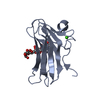
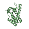
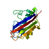
- Structure visualization
| Structure viewer | Molecule: MolmilJmol/JSmol |
|---|
- Downloads & links
-Download
| PDBx/mmCIF format | 3oea.cif.gz | 86.4 KB | Display | PDBx/mmCIF format |
|---|---|---|---|---|
| PDB format | pdb3oea.ent.gz | 64.1 KB | Display | PDB format |
| PDBx/mmJSON format | 3oea.json.gz | Tree view | PDBx/mmJSON format | |
| Others |  Other downloads Other downloads |
-Validation report
| Arichive directory | https://data.pdbj.org/pub/pdb/validation_reports/oe/3oeaftp://data.pdbj.org/pub/pdb/validation_reports/oe/3oea | HTTPS FTP |
|---|
-Related structure data
| Related structure data | 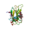 3oebC  2zexS C: citing same article ( S: Starting model for refinement |
|---|---|
| Similar structure data |


-Links
 PDBj
PDBj- Assembly
| Deposited unit | 
| ||||||||
|---|---|---|---|---|---|---|---|---|---|
| 1 | 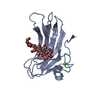
| ||||||||
| 2 | 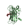
| ||||||||
| 3 | 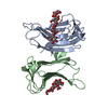
| ||||||||
| Unit cell |
|
-Components
| #1: Protein | Mass: 16050.784 Da / Num. of mol.: 2 / Fragment: UNP residues 614-756 / Mutation: Q121E Source method: isolated from a genetically manipulated source Source: (gene. exp.) Caldanaerobius polysaccharolyticus (bacteria)Gene: celA / Production host: #2: Polysaccharide | Source method: isolated from a genetically manipulated source #3: Chemical |   Mass: 40.078 Da / Num. of mol.: 2 / Source method: obtained synthetically / Formula: Ca Mass: 40.078 Da / Num. of mol.: 2 / Source method: obtained synthetically / Formula: Ca#4: Water | ChemComp-HOH / |  Mass: 18.015 Da / Num. of mol.: 497 / Source method: isolated from a natural source / Formula: H2O Mass: 18.015 Da / Num. of mol.: 497 / Source method: isolated from a natural source / Formula: H2O |
|---|
-Experimental details
-Experiment
| Experiment | Method: X-RAY DIFFRACTION / Number of used crystals: 1 |
|---|
- Sample preparation
| Crystal | Density Matthews: 2.11 Å3/Da / Density % sol: 41.77 % |
|---|---|
| Crystal grow | Temperature: 288 K / Method: vapor diffusion, hanging drop / pH: 8.3 Details: 30% PEG 3350, 100 mM Tris-HCl, 200 mM MgCl2, pH 8.3, VAPOR DIFFUSION, HANGING DROP, temperature 288K |
-Data collection
| Diffraction | Mean temperature: 100 K |
|---|---|
| Diffraction source | Source: SYNCHROTRON / Site: APS  / Beamline: 21-ID-D / Beamline: 21-ID-D |
| Detector | Type: MARMOSAIC 300 mm CCD / Detector: CCD / Date: Jul 15, 2010 |
| Radiation | Protocol: SINGLE WAVELENGTH / Monochromatic (M) / Laue (L): M / Scattering type: x-ray |
| Radiation wavelength | Relative weight: 1 |
| Reflection | Resolution: 1.35→25 Å / Num. all: 54672 / Num. obs: 54672 / % possible obs: 94.9 % / Observed criterion σ(F): 0 / Redundancy: 4.9 % / Rmerge(I) obs: 0.047 / Net I/σ(I): 45.9 |
| Reflection shell | Resolution: 1.35→1.37 Å / Redundancy: 4.7 % / Rmerge(I) obs: 0.145 / Mean I/σ(I) obs: 9.4 / Num. unique all: 2632 / % possible all: 91.5 |
- Processing
| Software |
| ||||||||||||||||||||||||||||||||||||||||||||||||||||||||||||||||||||||||||||||||
|---|---|---|---|---|---|---|---|---|---|---|---|---|---|---|---|---|---|---|---|---|---|---|---|---|---|---|---|---|---|---|---|---|---|---|---|---|---|---|---|---|---|---|---|---|---|---|---|---|---|---|---|---|---|---|---|---|---|---|---|---|---|---|---|---|---|---|---|---|---|---|---|---|---|---|---|---|---|---|---|---|---|
| Refinement | Method to determine structure: MOLECULAR REPLACEMENT Starting model: pdb entry 2ZEX Resolution: 1.35→25 Å / Cor.coef. Fo:Fc: 0.966 / Cor.coef. Fo:Fc free: 0.95 / SU B: 0.909 / SU ML: 0.039 / Cross valid method: THROUGHOUT / ESU R Free: 0.068 / Stereochemistry target values: MAXIMUM LIKELIHOOD / Details: HYDROGENS HAVE BEEN ADDED IN THE RIDING POSITIONS
| ||||||||||||||||||||||||||||||||||||||||||||||||||||||||||||||||||||||||||||||||
| Solvent computation | Ion probe radii: 0.8 Å / Shrinkage radii: 0.8 Å / VDW probe radii: 1.4 Å / Solvent model: BABINET MODEL WITH MASK | ||||||||||||||||||||||||||||||||||||||||||||||||||||||||||||||||||||||||||||||||
| Displacement parameters | Biso mean: 14.207 Å2
| ||||||||||||||||||||||||||||||||||||||||||||||||||||||||||||||||||||||||||||||||
| Refinement step | Cycle: LAST / Resolution: 1.35→25 Å
| ||||||||||||||||||||||||||||||||||||||||||||||||||||||||||||||||||||||||||||||||
| Refine LS restraints |
| ||||||||||||||||||||||||||||||||||||||||||||||||||||||||||||||||||||||||||||||||
| LS refinement shell | Resolution: 1.35→1.386 Å / Total num. of bins used: 20
|
