 Movie
Movie Controller

[English] 日本語
 Yorodumi
- PDB-1knk: Crystal Structure of 2-C-methyl-D-erythritol 2,4-cyclodiphosphate... -
+ Open data

- Basic information
| Entry | Database: PDB / ID: 1knk | ||||||
|---|---|---|---|---|---|---|---|
| Title | Crystal Structure of 2-C-methyl-D-erythritol 2,4-cyclodiphosphate Synthase (ispF) from E. coli involved in Mevalonate-Independent Isoprenoid Biosynthesis | ||||||
 Components Components | 2C-methyl-D-erythritol 2,4-cyclodiphosphate synthase | ||||||
 Keywords Keywords | METAL BINDING PROTEIN / isoprenoids / deoxyxylulose/methyl-erythritol-phosphate pathway / cyclodiphosphate / MEP / YgbB / ispF / MECDP / 2-C-methyl-D-erythritol-2 / 4-cyclodiphosphate synthase | ||||||
| Function / homology |  Function and homology information Function and homology information2-C-methyl-D-erythritol 2,4-cyclodiphosphate synthase / 2-C-methyl-D-erythritol 2,4-cyclodiphosphate synthase activity / isopentenyl diphosphate biosynthetic process, methylerythritol 4-phosphate pathway / terpenoid biosynthetic process / ubiquinone biosynthetic process / manganese ion binding / zinc ion binding / metal ion binding / identical protein binding Similarity search - Function | ||||||
| Biological species |  | ||||||
| Method |  X-RAY DIFFRACTION / SYNCHROTRON / Rigid body / Resolution: 2.8 Å X-RAY DIFFRACTION / SYNCHROTRON / Rigid body / Resolution: 2.8 Å | ||||||
 Authors Authors | Richard, S.B. / Ferrer, J.L. / Bowman, M.E. / Lillo, A.M. / Tetzlaff, C.N. / Cane, D.E. / Noel, J.P. | ||||||
 Citation Citation | Journal: J.Biol.Chem. / Year: 2002 Title: Structure and mechanism of 2-C-methyl-D-erythritol 2,4-cyclodiphosphate synthase. An enzyme in the mevalonate-independent isoprenoid biosynthetic pathway. Authors: Richard, S.B. / Ferrer, J.L. / Bowman, M.E. / Lillo, A.M. / Tetzlaff, C.N. / Cane, D.E. / Noel, J.P. #1: Journal: Nat.Struct.Biol. / Year: 2001Title: Structure of 4-diphosphocytidyl-2-C-methylerythritol Synthetase Involved in Mevalonate-Independent Isoprenoid Biosynthesis Authors: Richard, S.B. / Bowman, M.E. / Kwiatkowski, W. / Kang, I. / Chow, C. / Lillo, A.M. / Cane, D.E. / Noel, J.P. | ||||||
| History |
|
- Structure visualization
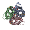
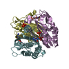
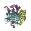
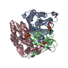
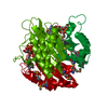
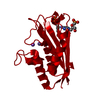
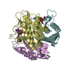
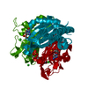
| Structure viewer | Molecule: MolmilJmol/JSmol |
|---|
- Downloads & links
-Download
| PDBx/mmCIF format | 1knk.cif.gz | 43.5 KB | Display | PDBx/mmCIF format |
|---|---|---|---|---|
| PDB format | pdb1knk.ent.gz | 30 KB | Display | PDB format |
| PDBx/mmJSON format | 1knk.json.gz | Tree view | PDBx/mmJSON format | |
| Others |  Other downloads Other downloads |
-Validation report
| Arichive directory | https://data.pdbj.org/pub/pdb/validation_reports/kn/1knkftp://data.pdbj.org/pub/pdb/validation_reports/kn/1knk | HTTPS FTP |
|---|
-Related structure data
| Related structure data |  1knjSC S: Starting model for refinement C: citing same article ( |
|---|---|
| Similar structure data |

-Links
 PDBj
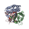
PDBj- Assembly
| Deposited unit | 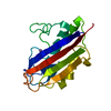
| |||||||||
|---|---|---|---|---|---|---|---|---|---|---|
| 1 | 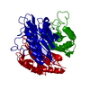
| |||||||||
| Unit cell |
| |||||||||
| Components on special symmetry positions |
| |||||||||
| Details | The biological assembly is a homotrimer generated from the monomer in the asymmetric unit by the operations: z,x,y and y,z,x |
-Components
| #1: Protein | Mass: 16920.531 Da / Num. of mol.: 1 Source method: isolated from a genetically manipulated source Source: (gene. exp.) |
|---|---|
| #2: Chemical | ChemComp-MN /   Mass: 54.938 Da / Num. of mol.: 1 / Source method: obtained synthetically / Formula: Mn Mass: 54.938 Da / Num. of mol.: 1 / Source method: obtained synthetically / Formula: Mn |
| #3: Water | ChemComp-HOH /  Mass: 18.015 Da / Num. of mol.: 11 / Source method: isolated from a natural source / Formula: H2O Mass: 18.015 Da / Num. of mol.: 11 / Source method: isolated from a natural source / Formula: H2O |
-Experimental details
-Experiment
| Experiment | Method: X-RAY DIFFRACTION / Number of used crystals: 1 |
|---|
- Sample preparation
| Crystal grow | Temperature: 294 K / Method: vapor diffusion, hanging drop / pH: 6.5 Details: (NH4)2SO4, PEG 400, NaI, PIPES, pH 6.5, VAPOR DIFFUSION, HANGING DROP, temperature 294K | ||||||||||||||||||||||||||||||||||||||||||
|---|---|---|---|---|---|---|---|---|---|---|---|---|---|---|---|---|---|---|---|---|---|---|---|---|---|---|---|---|---|---|---|---|---|---|---|---|---|---|---|---|---|---|---|
| Crystal grow | *PLUS Temperature: 4 ℃ | ||||||||||||||||||||||||||||||||||||||||||
| Components of the solutions | *PLUS
|
-Data collection
| Diffraction | Mean temperature: 100 K |
|---|---|
| Diffraction source | Source: SYNCHROTRON / Site: ESRF  / Beamline: ID14-4 / Wavelength: 0.9793 Å / Beamline: ID14-4 / Wavelength: 0.9793 Å |
| Detector | Type: ADSC QUANTUM 4 / Detector: CCD / Date: Oct 10, 2001 |
| Radiation | Monochromator: Si(111) OR Si(311) CRYSTALS, LN2 COOLED / Protocol: SINGLE WAVELENGTH / Monochromatic (M) / Laue (L): M / Scattering type: x-ray |
| Radiation wavelength | Wavelength: 0.9793 Å / Relative weight: 1 |
| Reflection | Resolution: 2.8→30 Å / Num. all: 24117 / Num. obs: 24117 / % possible obs: 93.6 % / Observed criterion σ(F): 1 / Observed criterion σ(I): 0 / Redundancy: 3.54 % / Biso Wilson estimate: 111.6 Å2 / Rsym value: 0.091 / Net I/σ(I): 15.7 |
| Reflection shell | Resolution: 2.8→2.9 Å / Redundancy: 3.3 % / Mean I/σ(I) obs: 5 / Num. unique all: 404 / Rsym value: 0.415 / % possible all: 96.9 |
| Reflection | *PLUS Highest resolution: 2.8 Å / Lowest resolution: 99 Å / Num. obs: 11929 / Redundancy: 3.55 % / Num. measured all: 41619 / Rmerge(I) obs: 0.091 |
| Reflection shell | *PLUS % possible obs: 96.9 % / Rmerge(I) obs: 0.415 |
- Processing
| Software |
| |||||||||||||||||||||||||
|---|---|---|---|---|---|---|---|---|---|---|---|---|---|---|---|---|---|---|---|---|---|---|---|---|---|---|
| Refinement | Method to determine structure: Rigid body Starting model: PDB ENTRY 1KNJ Resolution: 2.8→24.74 Å / Rfactor Rfree error: 0.008 / Data cutoff high absF: 623227.17 / Data cutoff low absF: 0 / Isotropic thermal model: RESTRAINED / Cross valid method: THROUGHOUT / σ(F): 0 / Stereochemistry target values: Engh & Huber
| |||||||||||||||||||||||||
| Solvent computation | Solvent model: FLAT MODEL / Bsol: 58.3984 Å2 / ksol: 0.369238 e/Å3 | |||||||||||||||||||||||||
| Displacement parameters | Biso mean: 72.3 Å2
| |||||||||||||||||||||||||
| Refine analyze |
| |||||||||||||||||||||||||
| Refinement step | Cycle: LAST / Resolution: 2.8→24.74 Å
| |||||||||||||||||||||||||
| Refine LS restraints |
| |||||||||||||||||||||||||
| LS refinement shell | Resolution: 2.8→2.98 Å / Rfactor Rfree error: 0.027 / Total num. of bins used: 6
| |||||||||||||||||||||||||
| Xplor file |
| |||||||||||||||||||||||||
| Refinement | *PLUS % reflection Rfree: 5 % / Rfactor Rfree: 0.253 / Rfactor Rwork: 0.243 | |||||||||||||||||||||||||
| Solvent computation | *PLUS | |||||||||||||||||||||||||
| Displacement parameters | *PLUS | |||||||||||||||||||||||||
| Refine LS restraints | *PLUS
| |||||||||||||||||||||||||
| LS refinement shell | *PLUS Rfactor Rfree: 0.367 / Rfactor Rwork: 0.366 |
