 Movie
Movie Controller

[English] 日本語
 Yorodumi
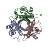
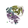
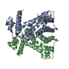
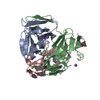
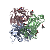
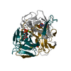
- PDB-3gwm: Crystal structure of the holo-[Acyl-Carrier-Protein] Synthase (AC... -
+ Open data

- Basic information
| Entry | Database: PDB / ID: 3gwm | ||||||
|---|---|---|---|---|---|---|---|
| Title | Crystal structure of the holo-[Acyl-Carrier-Protein] Synthase (ACPS) from Mycobacterium smegmatis | ||||||
 Components Components | Holo-[acyl-carrier-protein] synthase | ||||||
 Keywords Keywords | TRANSFERASE / Homo-trimer / 9-stand pseudo beta barrel protein / Cytoplasm / Fatty acid biosynthesis / Lipid synthesis / Magnesium / Metal-binding | ||||||
| Function / homology |  Function and homology information Function and homology informationholo-[acyl-carrier-protein] synthase / holo-[acyl-carrier-protein] synthase activity / fatty acid biosynthetic process / magnesium ion binding / cytoplasm Similarity search - Function | ||||||
| Biological species |  Mycobacterium smegmatis (bacteria) Mycobacterium smegmatis (bacteria) | ||||||
| Method |  X-RAY DIFFRACTION / SYNCHROTRON / SIRAS / Resolution: 1.7 Å X-RAY DIFFRACTION / SYNCHROTRON / SIRAS / Resolution: 1.7 Å | ||||||
 Authors Authors | Poulsen, C. / Wilmanns, M. / Song, Y.H. | ||||||
 Citation Citation | Journal: To be Published Title: Structure of the holo-[Acyl-Carrier-Protein] Synthase (ACPS) from Mycobacterium Authors: Poulsen, C. / Wilmanns, M. / Song, Y.H. | ||||||
| History |
|
- Structure visualization
| Structure viewer | Molecule: MolmilJmol/JSmol |
|---|
- Downloads & links
-Download
| PDBx/mmCIF format | 3gwm.cif.gz | 61.8 KB | Display | PDBx/mmCIF format |
|---|---|---|---|---|
| PDB format | pdb3gwm.ent.gz | 46 KB | Display | PDB format |
| PDBx/mmJSON format | 3gwm.json.gz | Tree view | PDBx/mmJSON format | |
| Others |  Other downloads Other downloads |
-Validation report
| Arichive directory | https://data.pdbj.org/pub/pdb/validation_reports/gw/3gwmftp://data.pdbj.org/pub/pdb/validation_reports/gw/3gwm | HTTPS FTP |
|---|
-Related structure data


-Links
 PDBj
PDBj- Assembly
| Deposited unit | 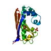
| ||||||||
|---|---|---|---|---|---|---|---|---|---|
| 1 | 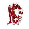
| ||||||||
| Unit cell |
| ||||||||
| Components on special symmetry positions |
|
-Components
| #1: Protein | Mass: 14054.824 Da / Num. of mol.: 1 / Source method: isolated from a natural source / Source: (natural) Mycobacterium smegmatis (bacteria) / Strain: MC2 155References: UniProt: A0R1H6, holo-[acyl-carrier-protein] synthase | ||
|---|---|---|---|
| #2: Chemical |   Mass: 96.063 Da / Num. of mol.: 3 / Source method: obtained synthetically / Formula: SO4 Mass: 96.063 Da / Num. of mol.: 3 / Source method: obtained synthetically / Formula: SO4#3: Water | ChemComp-HOH / |  Mass: 18.015 Da / Num. of mol.: 155 / Source method: isolated from a natural source / Formula: H2O Mass: 18.015 Da / Num. of mol.: 155 / Source method: isolated from a natural source / Formula: H2O |
-Experimental details
-Experiment
| Experiment | Method: X-RAY DIFFRACTION / Number of used crystals: 1 |
|---|
- Sample preparation
| Crystal | Density Matthews: 2.68 Å3/Da / Density % sol: 54.11 % |
|---|---|
| Crystal grow | Temperature: 297 K / Method: vapor diffusion, hanging drop / pH: 6.5 Details: 100mM Bis-Tris pH 6.5, 16% PEG 400, 200mM (NH4)SO4, VAPOR DIFFUSION, HANGING DROP, temperature 297K |
-Data collection
| Diffraction | Mean temperature: 100 K | ||||||||||||||||||||||||||||||||||||||||||||||||||||||||||||||||||||||||||||||||||||||||||||||||||||||||||||||||||||||||||||||
|---|---|---|---|---|---|---|---|---|---|---|---|---|---|---|---|---|---|---|---|---|---|---|---|---|---|---|---|---|---|---|---|---|---|---|---|---|---|---|---|---|---|---|---|---|---|---|---|---|---|---|---|---|---|---|---|---|---|---|---|---|---|---|---|---|---|---|---|---|---|---|---|---|---|---|---|---|---|---|---|---|---|---|---|---|---|---|---|---|---|---|---|---|---|---|---|---|---|---|---|---|---|---|---|---|---|---|---|---|---|---|---|---|---|---|---|---|---|---|---|---|---|---|---|---|---|---|---|
| Diffraction source | Source: SYNCHROTRON / Site: ESRF  / Beamline: ID23-1 / Wavelength: 1 Å / Beamline: ID23-1 / Wavelength: 1 Å | ||||||||||||||||||||||||||||||||||||||||||||||||||||||||||||||||||||||||||||||||||||||||||||||||||||||||||||||||||||||||||||||
| Detector | Type: ADSC QUANTUM 315r / Detector: CCD / Date: Apr 23, 2008 | ||||||||||||||||||||||||||||||||||||||||||||||||||||||||||||||||||||||||||||||||||||||||||||||||||||||||||||||||||||||||||||||
| Radiation | Protocol: SINGLE WAVELENGTH / Monochromatic (M) / Laue (L): M / Scattering type: x-ray | ||||||||||||||||||||||||||||||||||||||||||||||||||||||||||||||||||||||||||||||||||||||||||||||||||||||||||||||||||||||||||||||
| Radiation wavelength | Wavelength: 1 Å / Relative weight: 1 | ||||||||||||||||||||||||||||||||||||||||||||||||||||||||||||||||||||||||||||||||||||||||||||||||||||||||||||||||||||||||||||||
| Reflection | Number: 32734 / Rmerge(I) obs: 0.094 / D res high: 2.1 Å / Num. obs: 8718 / % possible obs: 99.4 | ||||||||||||||||||||||||||||||||||||||||||||||||||||||||||||||||||||||||||||||||||||||||||||||||||||||||||||||||||||||||||||||
| Diffraction reflection shell |
| ||||||||||||||||||||||||||||||||||||||||||||||||||||||||||||||||||||||||||||||||||||||||||||||||||||||||||||||||||||||||||||||
| Reflection | Resolution: 1.7→25 Å / Num. all: 16195 / Num. obs: 15966 / % possible obs: 98.6 % / Observed criterion σ(I): -3 / Redundancy: 3.1 % / Biso Wilson estimate: 26.967 Å2 / Rmerge(I) obs: 0.047 | ||||||||||||||||||||||||||||||||||||||||||||||||||||||||||||||||||||||||||||||||||||||||||||||||||||||||||||||||||||||||||||||
| Reflection shell | Resolution: 1.7→1.74 Å / Redundancy: 2.7 % / Rmerge(I) obs: 0.476 / Mean I/σ(I) obs: 2 / Num. measured obs: 2864 / Num. unique all: 1207 / Num. unique obs: 1080 / % possible all: 89.5 |
- Processing
| Software |
| |||||||||||||||||||||||||||||||||||||||||||||||||
|---|---|---|---|---|---|---|---|---|---|---|---|---|---|---|---|---|---|---|---|---|---|---|---|---|---|---|---|---|---|---|---|---|---|---|---|---|---|---|---|---|---|---|---|---|---|---|---|---|---|---|
| Refinement | Method to determine structure: SIRAS / Resolution: 1.7→21.851 Å / Occupancy max: 1 / Occupancy min: 0.44 / FOM work R set: 0.862 / SU ML: 0.09 / Cross valid method: THROUGHOUT / σ(F): 1.99 / Stereochemistry target values: ML
| |||||||||||||||||||||||||||||||||||||||||||||||||
| Solvent computation | Shrinkage radii: 0.9 Å / VDW probe radii: 1.11 Å / Solvent model: FLAT BULK SOLVENT MODEL / Bsol: 56.365 Å2 / ksol: 0.438 e/Å3 | |||||||||||||||||||||||||||||||||||||||||||||||||
| Displacement parameters | Biso max: 106.41 Å2 / Biso mean: 25.937 Å2 / Biso min: 7.07 Å2
| |||||||||||||||||||||||||||||||||||||||||||||||||
| Refinement step | Cycle: LAST / Resolution: 1.7→21.851 Å
| |||||||||||||||||||||||||||||||||||||||||||||||||
| Refine LS restraints |
| |||||||||||||||||||||||||||||||||||||||||||||||||
| LS refinement shell | Refine-ID: X-RAY DIFFRACTION / Total num. of bins used: 6
|
