 Movie
Movie Controller


+ Open data

- Basic information
| Entry | Database: PDB / ID: 1dup | ||||||
|---|---|---|---|---|---|---|---|
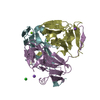
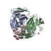
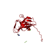
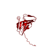
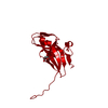
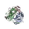
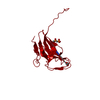
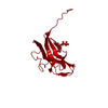
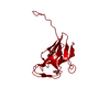
| Title | DEOXYURIDINE 5'-TRIPHOSPHATE NUCLEOTIDO HYDROLASE (D-UTPASE) | ||||||
 Components Components | DEOXYURIDINE 5'-TRIPHOSPHATE NUCLEOTIDOHYDROLASE | ||||||
 Keywords Keywords | HYDROLASE / NUCLEOTIDE METABOLISM | ||||||
| Function / homology |  Function and homology information Function and homology informationdUTP catabolic process / dUMP biosynthetic process / dUTP diphosphatase / dUTP diphosphatase activity / protein homotrimerization / magnesium ion binding / protein-containing complex / identical protein binding / cytosol Similarity search - Function | ||||||
| Biological species |  | ||||||
| Method |  X-RAY DIFFRACTION / Resolution: 1.9 Å X-RAY DIFFRACTION / Resolution: 1.9 Å | ||||||
 Authors Authors | Dauter, Z. / Wilson, K.S. / Larsson, G. / Nyman, P.O. / Cedergren, E. | ||||||
 Citation Citation | Journal: Nature / Year: 1992 Title: Crystal structure of a dUTPase. Authors: Cedergren-Zeppezauer, E.S. / Larsson, G. / Nyman, P.O. / Dauter, Z. / Wilson, K.S. | ||||||
| History |
|
- Structure visualization
| Structure viewer | Molecule: MolmilJmol/JSmol |
|---|
- Downloads & links
-Download
| PDBx/mmCIF format | 1dup.cif.gz | 40.9 KB | Display | PDBx/mmCIF format |
|---|---|---|---|---|
| PDB format | pdb1dup.ent.gz | 28.5 KB | Display | PDB format |
| PDBx/mmJSON format | 1dup.json.gz | Tree view | PDBx/mmJSON format | |
| Others |  Other downloads Other downloads |
-Validation report
| Arichive directory | https://data.pdbj.org/pub/pdb/validation_reports/du/1dupftp://data.pdbj.org/pub/pdb/validation_reports/du/1dup | HTTPS FTP |
|---|
-Related structure data
| Similar structure data |
|---|

-Links
 PDBj
PDBj
- Assembly
| Deposited unit | 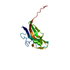
| |||||||||
|---|---|---|---|---|---|---|---|---|---|---|
| 1 | 
| |||||||||
| Unit cell |
| |||||||||
| Components on special symmetry positions |
| |||||||||
| Details | THE STRUCTURE IS BUILT-UP OF TIGHT TRIMERS AROUND THE CRYSTALLOGRAPHIC THREE-FOLD AXIS. MTRIX THE TRANSFORMATIONS PRESENTED ON MTRIX RECORDS BELOW DESCRIBE NON-CRYSTALLOGRAPHIC RELATIONSHIPS AMONG THE VARIOUS DOMAINS IN THIS ENTRY. APPLYING THE APPROPRIATE MTRIX TRANSFORMATION TO THE RESIDUES LISTED FIRST WILL YIELD APPROXIMATE COORDINATES FOR THE RESIDUES LISTED SECOND. APPLIED TO TRANSFORMED TO MTRIX RESIDUES RESIDUES RMSD M1 A 1 .. A 136 1 .. 136 M2 A 1 .. A 136 1 .. 136 THESE TRANSFORMATIONS WILL YIELD THE COORDINATES FOR THE TWO OTHER SUBUNITS OF THE TRIMER. |
-Components
| #1: Protein | Mass: 16302.615 Da / Num. of mol.: 1 / Source method: isolated from a natural source / Source: (natural) |
|---|---|
| #2: Water | ChemComp-HOH /  Mass: 18.015 Da / Num. of mol.: 123 / Source method: isolated from a natural source / Formula: H2O Mass: 18.015 Da / Num. of mol.: 123 / Source method: isolated from a natural source / Formula: H2O |
| Compound details | SECONDARY STRUCTURE BOUNDARIES HAVE BEEN DETERMINED USING SS PROGRAM (V.S.LAMZIN, EMBL HAMBURG) AS ...SECONDARY STRUCTURE BOUNDARIES |
-Experimental details
-Experiment
| Experiment | Method: X-RAY DIFFRACTION |
|---|
- Sample preparation
| Crystal | Density Matthews: 2.76 Å3/Da / Density % sol: 55.36 % | ||||||||||||||||||||||||||||||
|---|---|---|---|---|---|---|---|---|---|---|---|---|---|---|---|---|---|---|---|---|---|---|---|---|---|---|---|---|---|---|---|
| Crystal grow | *PLUS pH: 4.2 / Method: other | ||||||||||||||||||||||||||||||
| Components of the solutions | *PLUS
|
-Data collection
| Radiation | Scattering type: x-ray |
|---|---|
| Radiation wavelength | Relative weight: 1 |
| Reflection | *PLUS Highest resolution: 1.9 Å / Num. obs: 13640 / % possible obs: 96 % / Rmerge(I) obs: 0.069 |
- Processing
| Software |
| ||||||||||||||||||||||||||||||||||||||||||||||||||||||||||||||||||||||||||||||||||||
|---|---|---|---|---|---|---|---|---|---|---|---|---|---|---|---|---|---|---|---|---|---|---|---|---|---|---|---|---|---|---|---|---|---|---|---|---|---|---|---|---|---|---|---|---|---|---|---|---|---|---|---|---|---|---|---|---|---|---|---|---|---|---|---|---|---|---|---|---|---|---|---|---|---|---|---|---|---|---|---|---|---|---|---|---|---|
| Refinement | Resolution: 1.9→10 Å / σ(F): 0 /
| ||||||||||||||||||||||||||||||||||||||||||||||||||||||||||||||||||||||||||||||||||||
| Refinement step | Cycle: LAST / Resolution: 1.9→10 Å
| ||||||||||||||||||||||||||||||||||||||||||||||||||||||||||||||||||||||||||||||||||||
| Refine LS restraints |
|
