 Movie
Movie Controller

[English] 日本語
 Yorodumi
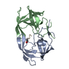
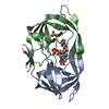
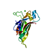
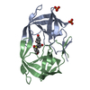
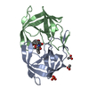
- PDB-3o7j: Crystal structure of 2-oxo-4-hydroxy-4-carboxy-5-ureidoimidazolin... -
+ Open data

- Basic information
| Entry | Database: PDB / ID: 3o7j | ||||||
|---|---|---|---|---|---|---|---|
| Title | Crystal structure of 2-oxo-4-hydroxy-4-carboxy-5-ureidoimidazoline decarboxylase from Klebsiella pneumoniae | ||||||
 Components Components | OHCU decarboxylase | ||||||
 Keywords Keywords | LYASE / decarboxylase | ||||||
| Function / homology |  Function and homology information Function and homology information2-oxo-4-hydroxy-4-carboxy-5-ureidoimidazoline decarboxylase / 2-oxo-4-hydroxy-4-carboxy-5-ureidoimidazoline decarboxylase activity / urate catabolic process / purine nucleobase metabolic process Similarity search - Function | ||||||
| Biological species |  Klebsiella pneumoniae subsp. pneumoniae (bacteria) Klebsiella pneumoniae subsp. pneumoniae (bacteria) | ||||||
| Method |  X-RAY DIFFRACTION / SYNCHROTRON / MOLECULAR REPLACEMENT / Resolution: 2 Å X-RAY DIFFRACTION / SYNCHROTRON / MOLECULAR REPLACEMENT / Resolution: 2 Å | ||||||
 Authors Authors | French, J.B. / Ealick, S.E. | ||||||
 Citation Citation | Journal: J.Biol.Chem. / Year: 2010 Title: Structural and Mechanistic Studies on Klebsiella pneumoniae 2-Oxo-4-hydroxy-4-carboxy-5-ureidoimidazoline Decarboxylase. Authors: French, J.B. / Ealick, S.E. | ||||||
| History |
|
- Structure visualization
| Structure viewer | Molecule: MolmilJmol/JSmol |
|---|
- Downloads & links
-Download
| PDBx/mmCIF format | 3o7j.cif.gz | 47.8 KB | Display | PDBx/mmCIF format |
|---|---|---|---|---|
| PDB format | pdb3o7j.ent.gz | 32.4 KB | Display | PDB format |
| PDBx/mmJSON format | 3o7j.json.gz | Tree view | PDBx/mmJSON format | |
| Others |  Other downloads Other downloads |
-Validation report
| Arichive directory | https://data.pdbj.org/pub/pdb/validation_reports/o7/3o7jftp://data.pdbj.org/pub/pdb/validation_reports/o7/3o7j | HTTPS FTP |
|---|
-Related structure data
| Related structure data | 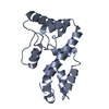 3o7hC 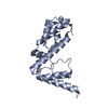 3o7iC 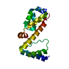 3o7kC 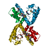 2q37S C: citing same article ( S: Starting model for refinement |
|---|---|
| Similar structure data |

-Links
 PDBj
PDBj- Assembly
| Deposited unit | 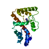
| |||||||||
|---|---|---|---|---|---|---|---|---|---|---|
| 1 |
| |||||||||
| Unit cell |
| |||||||||
| Components on special symmetry positions |
|
-Components
| #1: Protein | Mass: 20833.295 Da / Num. of mol.: 1 Source method: isolated from a genetically manipulated source Source: (gene. exp.) Klebsiella pneumoniae subsp. pneumoniae (bacteria)Strain: ATCC 700721 / Gene: KPN78578_16350, KPN_01665 / Production host: |
|---|---|
| #2: Chemical | ChemComp-2AL / 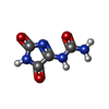  Mass: 156.100 Da / Num. of mol.: 1 / Source method: obtained synthetically / Formula: C4H4N4O3 Mass: 156.100 Da / Num. of mol.: 1 / Source method: obtained synthetically / Formula: C4H4N4O3 |
| #3: Water | ChemComp-HOH /  Mass: 18.015 Da / Num. of mol.: 89 / Source method: isolated from a natural source / Formula: H2O Mass: 18.015 Da / Num. of mol.: 89 / Source method: isolated from a natural source / Formula: H2O |
-Experimental details
-Experiment
| Experiment | Method: X-RAY DIFFRACTION / Number of used crystals: 1 |
|---|
- Sample preparation
| Crystal | Density Matthews: 2.18 Å3/Da / Density % sol: 43.5 % |
|---|---|
| Crystal grow | Temperature: 291 K / Method: vapor diffusion, hanging drop / pH: 6.5 Details: 22-26% PEG-8000, 0.25 M sodium acetate in pH 6.5 cacodylate, VAPOR DIFFUSION, HANGING DROP, temperature 291K |
-Data collection
| Diffraction | Mean temperature: 100 K |
|---|---|
| Diffraction source | Source: SYNCHROTRON / Site: CHESS  / Beamline: A1 / Wavelength: 0.987 Å / Beamline: A1 / Wavelength: 0.987 Å |
| Detector | Type: ADSC QUANTUM 210 / Detector: CCD / Date: Jan 1, 2010 |
| Radiation | Monochromator: Horizontal focusing 5.05 asymmetric cut Si(111) Protocol: SINGLE WAVELENGTH / Monochromatic (M) / Laue (L): M / Scattering type: x-ray |
| Radiation wavelength | Wavelength: 0.987 Å / Relative weight: 1 |
| Reflection | Resolution: 2→50 Å / Num. all: 12300 / Num. obs: 12100 / % possible obs: 98.6 % / Observed criterion σ(I): 2 / Redundancy: 2.4 % / Rsym value: 0.076 / Net I/σ(I): 16.5 |
| Reflection shell | Resolution: 2→2.03 Å / Redundancy: 2 % / Mean I/σ(I) obs: 2.6 / Num. unique all: 560 / Rsym value: 0.263 / % possible all: 94.1 |
- Processing
| Software |
| |||||||||||||||||||||||||||||||||||||||||||||||||||||||||||||||||
|---|---|---|---|---|---|---|---|---|---|---|---|---|---|---|---|---|---|---|---|---|---|---|---|---|---|---|---|---|---|---|---|---|---|---|---|---|---|---|---|---|---|---|---|---|---|---|---|---|---|---|---|---|---|---|---|---|---|---|---|---|---|---|---|---|---|---|
| Refinement | Method to determine structure: MOLECULAR REPLACEMENT Starting model: PDB ENTRY 2Q37 Resolution: 2→25.61 Å / Cor.coef. Fo:Fc: 0.939 / Cor.coef. Fo:Fc free: 0.915 / SU B: 4.809 / SU ML: 0.135 / Cross valid method: THROUGHOUT / ESU R Free: 0.171 / Stereochemistry target values: MAXIMUM LIKELIHOOD / Details: HYDROGENS HAVE BEEN ADDED IN THE RIDING POSITIONS
| |||||||||||||||||||||||||||||||||||||||||||||||||||||||||||||||||
| Solvent computation | Ion probe radii: 0.8 Å / Shrinkage radii: 0.8 Å / VDW probe radii: 1.4 Å / Solvent model: MASK | |||||||||||||||||||||||||||||||||||||||||||||||||||||||||||||||||
| Displacement parameters | Biso mean: 25.162 Å2
| |||||||||||||||||||||||||||||||||||||||||||||||||||||||||||||||||
| Refinement step | Cycle: LAST / Resolution: 2→25.61 Å
| |||||||||||||||||||||||||||||||||||||||||||||||||||||||||||||||||
| Refine LS restraints |
| |||||||||||||||||||||||||||||||||||||||||||||||||||||||||||||||||
| LS refinement shell | Resolution: 2→2.05 Å / Total num. of bins used: 20
|
