 Movie
Movie Controller


+ Open data

- Basic information
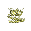
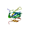
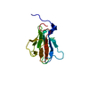
| Entry | Database: PDB / ID: 3o27 | ||||||
|---|---|---|---|---|---|---|---|
| Title | The crystal structure of C68 from the hybrid virus-plasmid pSSVx | ||||||
 Components Components | Putative uncharacterized protein | ||||||
 Keywords Keywords | DNA BINDING PROTEIN / swapped-hairpin fold / transcription factor | ||||||
| Function / homology | Pemi-like Protein 1; Chain: D / Pemi-like Protein 1; Chain: D - #10 / Antidote-toxin recognition MazE, bacterial antitoxin / SpoVT-AbrB domain / Ribbon / DNA binding / Mainly Beta / SpoVT-AbrB domain-containing protein Function and homology information Function and homology information | ||||||
| Biological species |   Sulfolobus islandicus (archaea) Sulfolobus islandicus (archaea) | ||||||
| Method |  X-RAY DIFFRACTION / SYNCHROTRON / SAD / Resolution: 2.8 Å X-RAY DIFFRACTION / SYNCHROTRON / SAD / Resolution: 2.8 Å | ||||||
 Authors Authors | D'Ambrosio, K. / De Simone, G. | ||||||
 Citation Citation | Journal: Biochem.J. / Year: 2011 Title: C68 from the Sulfolobus islandicus plasmid-virus pSSVx is a novel member of the AbrB-like transcription factor family. Authors: Contursi, P. / D'Ambrosio, K. / Pirone, L. / Pedone, E. / Aucelli, T. / She, Q. / De Simone, G. / Bartolucci, S. | ||||||
| History |
|
- Structure visualization
| Structure viewer | Molecule: MolmilJmol/JSmol |
|---|
- Downloads & links
-Download
| PDBx/mmCIF format | 3o27.cif.gz | 35.2 KB | Display | PDBx/mmCIF format |
|---|---|---|---|---|
| PDB format | pdb3o27.ent.gz | 25.3 KB | Display | PDB format |
| PDBx/mmJSON format | 3o27.json.gz | Tree view | PDBx/mmJSON format | |
| Others |  Other downloads Other downloads |
-Validation report
| Summary document | 3o27_validation.pdf.gz | 430.5 KB | Display | wwPDB validaton report |
|---|---|---|---|---|
| Full document | 3o27_full_validation.pdf.gz | 432.5 KB | Display | |
| Data in XML | 3o27_validation.xml.gz | 7.2 KB | Display | |
| Data in CIF | 3o27_validation.cif.gz | 8.9 KB | Display | |
| Arichive directory | https://data.pdbj.org/pub/pdb/validation_reports/o2/3o27ftp://data.pdbj.org/pub/pdb/validation_reports/o2/3o27 | HTTPS FTP |
-Related structure data
| Similar structure data |
|---|


-Links
 PDBj
PDBj- Assembly
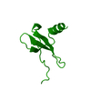
| Deposited unit | 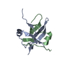
| ||||||||
|---|---|---|---|---|---|---|---|---|---|
| 1 |
| ||||||||
| Unit cell |
|
-Components
| #1: Protein | Mass: 7757.146 Da / Num. of mol.: 2 Source method: isolated from a genetically manipulated source Source: (gene. exp.) Sulfolobus islandicus (archaea) / Strain: REY15/4 / Gene: orfc68 / Plasmid: pET30 / Production host:  #2: Water | ChemComp-HOH / |  Mass: 18.015 Da / Num. of mol.: 38 / Source method: isolated from a natural source / Formula: H2O Mass: 18.015 Da / Num. of mol.: 38 / Source method: isolated from a natural source / Formula: H2O |
|---|
-Experimental details
-Experiment
| Experiment | Method: X-RAY DIFFRACTION / Number of used crystals: 1 |
|---|
- Sample preparation
| Crystal | Density Matthews: 3.36 Å3/Da / Density % sol: 63.36 % |
|---|---|
| Crystal grow | Temperature: 298 K / Method: vapor diffusion, hanging drop / pH: 3.6 Details: 1.6 M sodium formate, 0.1 M Sodium acetate, pH 3.6, VAPOR DIFFUSION, HANGING DROP, temperature 298K |
-Data collection
| Diffraction | Mean temperature: 100 K |
|---|---|
| Diffraction source | Source: SYNCHROTRON / Site: ELETTRA  / Beamline: 5.2R / Wavelength: 1 Å / Beamline: 5.2R / Wavelength: 1 Å |
| Detector | Type: MAR CCD 165 mm / Detector: CCD / Date: Oct 29, 2008 |
| Radiation | Monochromator: Graphite / Protocol: SINGLE WAVELENGTH / Monochromatic (M) / Laue (L): M / Scattering type: x-ray |
| Radiation wavelength | Wavelength: 1 Å / Relative weight: 1 |
| Reflection | Resolution: 2.8→50 Å / Num. all: 5275 / Num. obs: 5275 / % possible obs: 100 % / Redundancy: 9.6 % / Rmerge(I) obs: 0.059 / Net I/σ(I): 0.393 |
| Reflection shell | Resolution: 2.8→2.9 Å / Redundancy: 9 % / Rmerge(I) obs: 0.444 / Mean I/σ(I) obs: 5.3 / Num. unique all: 528 / % possible all: 99.8 |
- Processing
| Software |
| ||||||||||||||||||||
|---|---|---|---|---|---|---|---|---|---|---|---|---|---|---|---|---|---|---|---|---|---|
| Refinement | Method to determine structure: SAD / Resolution: 2.8→50 Å / σ(F): 0 / Stereochemistry target values: Engh & Huber
| ||||||||||||||||||||
| Refinement step | Cycle: LAST / Resolution: 2.8→50 Å
| ||||||||||||||||||||
| Refine LS restraints |
| ||||||||||||||||||||
| LS refinement shell | Resolution: 2.8→2.93 Å /
|
