 Movie
Movie Controller

[English] 日本語
 Yorodumi
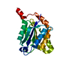
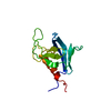
- PDB-3hqc: Crystal structure of Phosphotyrosine-binding domain from the Huma... -
+ Open data

- Basic information
| Entry | Database: PDB / ID: 3hqc | ||||||
|---|---|---|---|---|---|---|---|
| Title | Crystal structure of Phosphotyrosine-binding domain from the Human Tensin-like C1 domain-containing phosphatase (TENC1) | ||||||
 Components Components | Tensin-like C1 domain-containing phosphatase | ||||||
 Keywords Keywords | HYDROLASE / Human Tensin-like C1 domain-containing phosphatase / TENC1 / Phosphotyrosine Binding Domain / PTB / TNS2 / KIAA1075 / STRUCTURAL GENOMICS / PSI-2 / PROTEIN STRUCTURE INITIATIVE / NEW YORK SGX Research Center for Structural Genomics / NYSGXRC / Cell junction / Cell membrane / Membrane / Metal-binding / Phorbol-ester binding / Phosphoprotein / Protein phosphatase / SH2 domain / Zinc-finger | ||||||
| Function / homology |  Function and homology information Function and homology informationmulticellular organismal-level homeostasis / collagen metabolic process / cellular homeostasis / response to muscle activity / peptidyl-tyrosine dephosphorylation / protein-tyrosine-phosphatase / negative regulation of insulin receptor signaling pathway / protein tyrosine phosphatase activity / kidney development / multicellular organism growth ...multicellular organismal-level homeostasis / collagen metabolic process / cellular homeostasis / response to muscle activity / peptidyl-tyrosine dephosphorylation / protein-tyrosine-phosphatase / negative regulation of insulin receptor signaling pathway / protein tyrosine phosphatase activity / kidney development / multicellular organism growth / kinase binding / negative regulation of cell population proliferation / focal adhesion / lipid binding / zinc ion binding / identical protein binding / plasma membrane / cytoplasm Similarity search - Function | ||||||
| Biological species |  Homo sapiens (human) Homo sapiens (human) | ||||||
| Method |  X-RAY DIFFRACTION / SYNCHROTRON / MOLECULAR REPLACEMENT / Resolution: 1.8 Å X-RAY DIFFRACTION / SYNCHROTRON / MOLECULAR REPLACEMENT / Resolution: 1.8 Å | ||||||
 Authors Authors | Sampathkumar, P. / Romero, R. / Wasserman, S. / Do, J. / Dickey, M. / Bain, K. / Gheyi, T. / Klemke, R. / Atwell, S. / Sauder, J.M. ...Sampathkumar, P. / Romero, R. / Wasserman, S. / Do, J. / Dickey, M. / Bain, K. / Gheyi, T. / Klemke, R. / Atwell, S. / Sauder, J.M. / Burley, S.K. / New York SGX Research Center for Structural Genomics (NYSGXRC) | ||||||
 Citation Citation | Journal: To be Published Title: Crystal structure of Phosphotyrosine-binding domain from the Human Tensin-like C1 domain-containing phosphatase (TENC1) Authors: Sampathkumar, P. / Romero, R. / Wasserman, S. / Do, J. / Dickey, M. / Bain, K. / Gheyi, T. / Klemke, R. / Atwell, S. / Sauder, J.M. / Burley, S.K. | ||||||
| History |
|
- Structure visualization
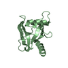
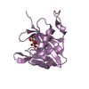
| Structure viewer | Molecule: MolmilJmol/JSmol |
|---|
- Downloads & links
-Download
| PDBx/mmCIF format | 3hqc.cif.gz | 46.2 KB | Display | PDBx/mmCIF format |
|---|---|---|---|---|
| PDB format | pdb3hqc.ent.gz | 31.5 KB | Display | PDB format |
| PDBx/mmJSON format | 3hqc.json.gz | Tree view | PDBx/mmJSON format | |
| Others |  Other downloads Other downloads |
-Validation report
| Arichive directory | https://data.pdbj.org/pub/pdb/validation_reports/hq/3hqcftp://data.pdbj.org/pub/pdb/validation_reports/hq/3hqc | HTTPS FTP |
|---|
-Related structure data
| Related structure data | 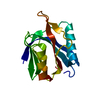 1wvhS S: Starting model for refinement |
|---|---|
| Similar structure data | |
| Other databases |


-Links
 PDBj
PDBj


- Assembly
| Deposited unit | 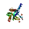
| ||||||||
|---|---|---|---|---|---|---|---|---|---|
| 1 |
| ||||||||
| Unit cell |
|
-Components
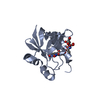
| #1: Protein | Mass: 17310.248 Da / Num. of mol.: 1 Fragment: Phosphotyrosine Binding Domain, PTB, residues 1264-1409 Source method: isolated from a genetically manipulated source Source: (gene. exp.) Homo sapiens (human) / Gene: KIAA1075, TENC1, TNS2 / Plasmid: BC-pSGX3(BC); modified pET26b / Production host:  References: UniProt: Q63HR2, Hydrolases; Acting on ester bonds; Phosphoric-monoester hydrolases | ||||||||
|---|---|---|---|---|---|---|---|---|---|
| #2: Chemical |   Mass: 96.063 Da / Num. of mol.: 3 / Source method: obtained synthetically / Formula: SO4 Mass: 96.063 Da / Num. of mol.: 3 / Source method: obtained synthetically / Formula: SO4#3: Chemical |   Mass: 59.044 Da / Num. of mol.: 2 / Source method: obtained synthetically / Formula: C2H3O2 Mass: 59.044 Da / Num. of mol.: 2 / Source method: obtained synthetically / Formula: C2H3O2#4: Chemical | ChemComp-GOL / |   Mass: 92.094 Da / Num. of mol.: 1 / Source method: obtained synthetically / Formula: C3H8O3 Mass: 92.094 Da / Num. of mol.: 1 / Source method: obtained synthetically / Formula: C3H8O3#5: Water | ChemComp-HOH / |  Mass: 18.015 Da / Num. of mol.: 79 / Source method: isolated from a natural source / Formula: H2O Mass: 18.015 Da / Num. of mol.: 79 / Source method: isolated from a natural source / Formula: H2OHas protein modification | Y | |
-Experimental details
-Experiment
| Experiment | Method: X-RAY DIFFRACTION / Number of used crystals: 1 |
|---|
- Sample preparation
| Crystal | Density Matthews: 3.58 Å3/Da / Density % sol: 65.64 % |
|---|---|
| Crystal grow | Temperature: 294 K / Method: vapor diffusion, sitting drop / pH: 4 Details: 100mM Sodium Acetate pH 4 + 2% Glycerol + 2000mM Ammonium Sulfate, VAPOR DIFFUSION, SITTING DROP, temperature 294K |
-Data collection
| Diffraction | Mean temperature: 100 K |
|---|---|
| Diffraction source | Source: SYNCHROTRON / Site: APS  / Beamline: 31-ID / Wavelength: 0.9793 Å / Beamline: 31-ID / Wavelength: 0.9793 Å |
| Detector | Type: MAR CCD 165 mm / Detector: CCD / Date: Feb 16, 2009 |
| Radiation | Monochromator: diamond / Protocol: SINGLE WAVELENGTH / Monochromatic (M) / Laue (L): M / Scattering type: x-ray |
| Radiation wavelength | Wavelength: 0.9793 Å / Relative weight: 1 |
| Reflection | Resolution: 1.8→47.14 Å / Num. obs: 23089 / % possible obs: 100 % / Redundancy: 10.9 % / Biso Wilson estimate: 25.92 Å2 / Rsym value: 0.118 / Net I/σ(I): 8.3 |
| Reflection shell | Resolution: 1.8→1.9 Å / Redundancy: 10.7 % / Mean I/σ(I) obs: 3 / Num. unique all: 3337 / Rsym value: 0.637 / % possible all: 100 |
- Processing
| Software |
| |||||||||||||||||||||||||||||||||||||||||||||||||||||||||||||||||||||||||||||||||||||||||||||||||||||||||||||||||||||||||||||
|---|---|---|---|---|---|---|---|---|---|---|---|---|---|---|---|---|---|---|---|---|---|---|---|---|---|---|---|---|---|---|---|---|---|---|---|---|---|---|---|---|---|---|---|---|---|---|---|---|---|---|---|---|---|---|---|---|---|---|---|---|---|---|---|---|---|---|---|---|---|---|---|---|---|---|---|---|---|---|---|---|---|---|---|---|---|---|---|---|---|---|---|---|---|---|---|---|---|---|---|---|---|---|---|---|---|---|---|---|---|---|---|---|---|---|---|---|---|---|---|---|---|---|---|---|---|---|
| Refinement | Method to determine structure: MOLECULAR REPLACEMENT Starting model: Poly-Alanine model of the PDB entry 1WVH Resolution: 1.8→26.16 Å / Cor.coef. Fo:Fc: 0.952 / Cor.coef. Fo:Fc free: 0.936 / Occupancy max: 1 / Occupancy min: 0.25 / SU B: 2.5 / SU ML: 0.078 / Cross valid method: THROUGHOUT / σ(F): 0 / ESU R: 0.105 / ESU R Free: 0.105 Stereochemistry target values: MAXIMUM LIKELIHOOD WITH PHASES Details: There are residual postive electron density features which could not be assigned unambigously as protein or buffer components.
| |||||||||||||||||||||||||||||||||||||||||||||||||||||||||||||||||||||||||||||||||||||||||||||||||||||||||||||||||||||||||||||
| Solvent computation | Ion probe radii: 0.8 Å / Shrinkage radii: 0.8 Å / VDW probe radii: 1.4 Å / Solvent model: BABINET MODEL WITH MASK | |||||||||||||||||||||||||||||||||||||||||||||||||||||||||||||||||||||||||||||||||||||||||||||||||||||||||||||||||||||||||||||
| Displacement parameters | Biso max: 79.88 Å2 / Biso mean: 33.104 Å2 / Biso min: 17.95 Å2
| |||||||||||||||||||||||||||||||||||||||||||||||||||||||||||||||||||||||||||||||||||||||||||||||||||||||||||||||||||||||||||||
| Refinement step | Cycle: LAST / Resolution: 1.8→26.16 Å
| |||||||||||||||||||||||||||||||||||||||||||||||||||||||||||||||||||||||||||||||||||||||||||||||||||||||||||||||||||||||||||||
| Refine LS restraints |
| |||||||||||||||||||||||||||||||||||||||||||||||||||||||||||||||||||||||||||||||||||||||||||||||||||||||||||||||||||||||||||||
| LS refinement shell | Resolution: 1.8→1.847 Å / Total num. of bins used: 20
|
