 Movie
Movie Controller

[English] 日本語
 Yorodumi
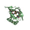
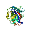
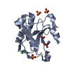
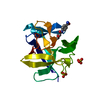
- PDB-1fhw: Structure of the pleckstrin homology domain from GRP1 in complex ... -
+ Open data

- Basic information
| Entry | Database: PDB / ID: 1fhw | ||||||
|---|---|---|---|---|---|---|---|
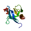
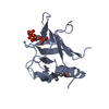
| Title | Structure of the pleckstrin homology domain from GRP1 in complex with inositol(1,3,4,5,6)pentakisphosphate | ||||||
 Components Components | GUANINE NUCLEOTIDE EXCHANGE FACTOR AND INTEGRIN BINDING PROTEIN HOMOLOG GRP1 | ||||||
 Keywords Keywords | SIGNALING PROTEIN / PLECKSTRIN / 3-PHOSPHOINOSITIDES / INOSITOL TETRAKISPHOSPHATE SIGNAL TRANSDUCTION PROTEIN / GUANINE NUCLEOTIDE EXCHANGE FACTOR | ||||||
| Function / homology |  Function and homology information Function and homology informationIntra-Golgi traffic / establishment of epithelial cell polarity / Golgi vesicle transport / regulation of ARF protein signal transduction / phosphatidylinositol-3,4,5-trisphosphate binding / bicellular tight junction / ruffle / positive regulation of cell adhesion / guanyl-nucleotide exchange factor activity / adherens junction ...Intra-Golgi traffic / establishment of epithelial cell polarity / Golgi vesicle transport / regulation of ARF protein signal transduction / phosphatidylinositol-3,4,5-trisphosphate binding / bicellular tight junction / ruffle / positive regulation of cell adhesion / guanyl-nucleotide exchange factor activity / adherens junction / nucleoplasm / plasma membrane / cytoplasm / cytosol Similarity search - Function | ||||||
| Biological species |  | ||||||
| Method |  X-RAY DIFFRACTION / SYNCHROTRON / MIR / Resolution: 1.9 Å X-RAY DIFFRACTION / SYNCHROTRON / MIR / Resolution: 1.9 Å | ||||||
 Authors Authors | Ferguson, K.M. / Kavran, J.M. / Sankaran, V.G. / Fournier, E. / Isakoff, S.J. / Skolnik, E.Y. / Lemmon, M.A. | ||||||
 Citation Citation | Journal: Mol.Cell / Year: 2000 Title: Structural basis for discrimination of 3-phosphoinositides by pleckstrin homology domains Authors: Ferguson, K.M. / Kavran, J.M. / Sankaran, V.G. / Fournier, E. / Isakoff, S.J. / Skolnik, E.Y. / Lemmon, M.A. | ||||||
| History |
|
- Structure visualization
| Structure viewer | Molecule: MolmilJmol/JSmol |
|---|
- Downloads & links
-Download
| PDBx/mmCIF format | 1fhw.cif.gz | 68.9 KB | Display | PDBx/mmCIF format |
|---|---|---|---|---|
| PDB format | pdb1fhw.ent.gz | 50.1 KB | Display | PDB format |
| PDBx/mmJSON format | 1fhw.json.gz | Tree view | PDBx/mmJSON format | |
| Others |  Other downloads Other downloads |
-Validation report
| Arichive directory | https://data.pdbj.org/pub/pdb/validation_reports/fh/1fhwftp://data.pdbj.org/pub/pdb/validation_reports/fh/1fhw | HTTPS FTP |
|---|
-Related structure data


-Links
 PDBj
PDBj
- Assembly
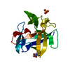
| Deposited unit | 
| ||||||||
|---|---|---|---|---|---|---|---|---|---|
| 1 | 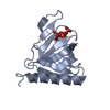
| ||||||||
| 2 | 
| ||||||||
| 3 |
| ||||||||
| Unit cell |
| ||||||||
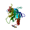
| Details | The biological assembly is a monomer constructed from chain A or chain B |
-Components
| #1: Protein | Mass: 15219.406 Da / Num. of mol.: 2 / Fragment: PLECKSTRIN HOMOLOGY DOMAIN Source method: isolated from a genetically manipulated source Details: COMPLEX WITH INOSITOL (1,3,4,5,6)-PENTAKISPHOSPHATE Source: (gene. exp.)  #2: Chemical | 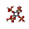  Mass: 580.055 Da / Num. of mol.: 2 / Source method: obtained synthetically / Formula: C6H17O21P5 Mass: 580.055 Da / Num. of mol.: 2 / Source method: obtained synthetically / Formula: C6H17O21P5#3: Chemical | ChemComp-SO4 / |   Mass: 96.063 Da / Num. of mol.: 1 / Source method: obtained synthetically / Formula: SO4 Mass: 96.063 Da / Num. of mol.: 1 / Source method: obtained synthetically / Formula: SO4#4: Water | ChemComp-HOH / |  Mass: 18.015 Da / Num. of mol.: 164 / Source method: isolated from a natural source / Formula: H2O Mass: 18.015 Da / Num. of mol.: 164 / Source method: isolated from a natural source / Formula: H2OHas protein modification | Y | |
|---|
-Experimental details
-Experiment
| Experiment | Method: X-RAY DIFFRACTION / Number of used crystals: 1 |
|---|
- Sample preparation
| Crystal | Density Matthews: 2.11 Å3/Da / Density % sol: 29.5 % | ||||||||||||||||||||||||||||||||||||||||||
|---|---|---|---|---|---|---|---|---|---|---|---|---|---|---|---|---|---|---|---|---|---|---|---|---|---|---|---|---|---|---|---|---|---|---|---|---|---|---|---|---|---|---|---|
| Crystal grow | Temperature: 291 K / Method: vapor diffusion, hanging drop / pH: 5 Details: PEG 8000, SODIUM ACETATE, AMMONIUM SULFATE, pH 5.00, VAPOR DIFFUSION, HANGING DROP, temperature 291K | ||||||||||||||||||||||||||||||||||||||||||
| Crystal grow | *PLUS Temperature: 21 ℃ / Method: vapor diffusion, hanging drop / Details: used microseeding | ||||||||||||||||||||||||||||||||||||||||||
| Components of the solutions | *PLUS
|
-Data collection
| Diffraction | Mean temperature: 100 K |
|---|---|
| Diffraction source | Source: SYNCHROTRON / Site: NSLS  / Beamline: X25 / Wavelength: 1 / Beamline: X25 / Wavelength: 1 |
| Detector | Type: BRANDEIS - B4 / Detector: CCD / Date: Feb 10, 1999 |
| Radiation | Protocol: SINGLE WAVELENGTH / Monochromatic (M) / Laue (L): M / Scattering type: x-ray |
| Radiation wavelength | Wavelength: 1 Å / Relative weight: 1 |
| Reflection | Resolution: 1.9→50 Å / Num. all: 53932 / Num. obs: 18374 / % possible obs: 95.2 % / Observed criterion σ(F): 0 / Observed criterion σ(I): 0 / Redundancy: 2.9 % / Biso Wilson estimate: 15 Å2 / Rmerge(I) obs: 0.05 / Rsym value: 0.05 / Net I/σ(I): 20 |
| Reflection shell | Resolution: 1.9→1.96 Å / Redundancy: 2.5 % / Rmerge(I) obs: 0.08 / Mean I/σ(I) obs: 10.7 / Rsym value: 0.08 / % possible all: 80.5 |
| Reflection | *PLUS Highest resolution: 1.9 Å / Lowest resolution: 50 Å / Observed criterion σ(I): 0 / Num. measured all: 53932 |
| Reflection shell | *PLUS % possible obs: 80.5 % |
- Processing
| Software |
| ||||||||||||||||||||||||||||||||||||||||
|---|---|---|---|---|---|---|---|---|---|---|---|---|---|---|---|---|---|---|---|---|---|---|---|---|---|---|---|---|---|---|---|---|---|---|---|---|---|---|---|---|---|
| Refinement | Method to determine structure: MIR / Resolution: 1.9→46.83 Å / Rfactor Rfree error: 0.006 / Data cutoff high absF: 1114520.05 / Data cutoff low absF: 0 / Isotropic thermal model: RESTRAINED / Cross valid method: THROUGHOUT / σ(F): 0 / σ(I): 0 / Stereochemistry target values: Engh & Huber
| ||||||||||||||||||||||||||||||||||||||||
| Solvent computation | Solvent model: FLAT MODEL / Bsol: 57.4 Å2 / ksol: 0.373 e/Å3 | ||||||||||||||||||||||||||||||||||||||||
| Displacement parameters | Biso mean: 27.3 Å2
| ||||||||||||||||||||||||||||||||||||||||
| Refine analyze |
| ||||||||||||||||||||||||||||||||||||||||
| Refinement step | Cycle: LAST / Resolution: 1.9→46.83 Å
| ||||||||||||||||||||||||||||||||||||||||
| Refine LS restraints |
| ||||||||||||||||||||||||||||||||||||||||
| LS refinement shell | Resolution: 1.9→2.02 Å / Rfactor Rfree error: 0.019 / Total num. of bins used: 6
| ||||||||||||||||||||||||||||||||||||||||
| Xplor file |
| ||||||||||||||||||||||||||||||||||||||||
| Software | *PLUS Name: CNS / Version: 0.9 / Classification: refinement | ||||||||||||||||||||||||||||||||||||||||
| Refinement | *PLUS Lowest resolution: 500 Å / σ(F): 0 / % reflection Rfree: 9.8 % / Rfactor Rfree: 0.27 | ||||||||||||||||||||||||||||||||||||||||
| Solvent computation | *PLUS | ||||||||||||||||||||||||||||||||||||||||
| Displacement parameters | *PLUS Biso mean: 27.3 Å2 | ||||||||||||||||||||||||||||||||||||||||
| Refine LS restraints | *PLUS
| ||||||||||||||||||||||||||||||||||||||||
| LS refinement shell | *PLUS Rfactor Rfree: 0.284 / % reflection Rfree: 10.2 % / Rfactor Rwork: 0.229 |
