 Movie
Movie Controller


+ Open data

- Basic information
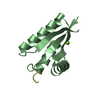
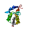
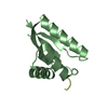
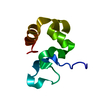
| Entry | Database: PDB / ID: 3o1f | ||||||
|---|---|---|---|---|---|---|---|
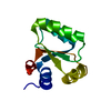
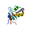
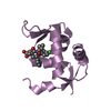
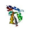
| Title | P1 crystal form of E. coli ClpS at 1.4 A resolution | ||||||
 Components Components | (ATP-dependent Clp protease adapter protein clpS) x 2 | ||||||
 Keywords Keywords | HYDROLASE / adaptor | ||||||
| Function / homology |  Function and homology information Function and homology informationmolecular function inhibitor activity / protein catabolic process / peptidase activity / protein-folding chaperone binding / response to heat / proteolysis Similarity search - Function | ||||||
| Biological species |  | ||||||
| Method |  X-RAY DIFFRACTION / MOLECULAR REPLACEMENT / Resolution: 1.4 Å X-RAY DIFFRACTION / MOLECULAR REPLACEMENT / Resolution: 1.4 Å | ||||||
 Authors Authors | Roman-Hernandez, G. / Hou, J.Y. / Grant, R.A. / Sauer, R.T. / Baker, T.A. | ||||||
 Citation Citation | Journal: Mol.Cell / Year: 2011 Title: The ClpS Adaptor Mediates Staged Delivery of N-End Rule Substrates to the AAA+ ClpAP Protease. Authors: Roman-Hernandez, G. / Hou, J.Y. / Grant, R.A. / Sauer, R.T. / Baker, T.A. | ||||||
| History |
|
- Structure visualization
| Structure viewer | Molecule: MolmilJmol/JSmol |
|---|
- Downloads & links
-Download
| PDBx/mmCIF format | 3o1f.cif.gz | 82.3 KB | Display | PDBx/mmCIF format |
|---|---|---|---|---|
| PDB format | pdb3o1f.ent.gz | 61.7 KB | Display | PDB format |
| PDBx/mmJSON format | 3o1f.json.gz | Tree view | PDBx/mmJSON format | |
| Others |  Other downloads Other downloads |
-Validation report
| Summary document | 3o1f_validation.pdf.gz | 406.1 KB | Display | wwPDB validaton report |
|---|---|---|---|---|
| Full document | 3o1f_full_validation.pdf.gz | 406.1 KB | Display | |
| Data in XML | 3o1f_validation.xml.gz | 15.3 KB | Display | |
| Data in CIF | 3o1f_validation.cif.gz | 22.1 KB | Display | |
| Arichive directory | https://data.pdbj.org/pub/pdb/validation_reports/o1/3o1fftp://data.pdbj.org/pub/pdb/validation_reports/o1/3o1f | HTTPS FTP |
-Related structure data


-Links
 PDBj
PDBj

- Assembly
| Deposited unit | 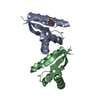
| ||||||||
|---|---|---|---|---|---|---|---|---|---|
| 1 | 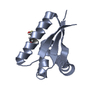
| ||||||||
| 2 | 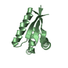
| ||||||||
| Unit cell |
|
-Components
| #1: Protein | Mass: 9305.767 Da / Num. of mol.: 1 / Fragment: unp residues 26-106 Source method: isolated from a genetically manipulated source Source: (gene. exp.) |
|---|---|
| #2: Protein | Mass: 9289.767 Da / Num. of mol.: 1 / Fragment: unp residues 26-106 Source method: isolated from a genetically manipulated source Source: (gene. exp.) |
| #3: Water | ChemComp-HOH /  Mass: 18.015 Da / Num. of mol.: 402 / Source method: isolated from a natural source / Formula: H2O Mass: 18.015 Da / Num. of mol.: 402 / Source method: isolated from a natural source / Formula: H2O |
| Has protein modification | Y |
-Experimental details
-Experiment
| Experiment | Method: X-RAY DIFFRACTION / Number of used crystals: 1 |
|---|
- Sample preparation
| Crystal | Density Matthews: 2.02 Å3/Da / Density % sol: 39.13 % |
|---|---|
| Crystal grow | Temperature: 300 K / Method: vapor diffusion, hanging drop / pH: 7 Details: 0.2M Ammonium Formate, 20% PEG 3350, VAPOR DIFFUSION, HANGING DROP, pH 7, temperature 300K |
-Data collection
| Diffraction | Mean temperature: 100 K | |||||||||||||||||||||||||||||||||||||||||||||||||||||||||||||||||||||||||||||
|---|---|---|---|---|---|---|---|---|---|---|---|---|---|---|---|---|---|---|---|---|---|---|---|---|---|---|---|---|---|---|---|---|---|---|---|---|---|---|---|---|---|---|---|---|---|---|---|---|---|---|---|---|---|---|---|---|---|---|---|---|---|---|---|---|---|---|---|---|---|---|---|---|---|---|---|---|---|---|
| Diffraction source | Source: ROTATING ANODE / Type: RIGAKU MICROMAX-007 HF / Wavelength: 1.5418 Å | |||||||||||||||||||||||||||||||||||||||||||||||||||||||||||||||||||||||||||||
| Detector | Type: RIGAKU RAXIS IV / Detector: IMAGE PLATE / Date: Jan 19, 2010 / Details: Varimax-HR | |||||||||||||||||||||||||||||||||||||||||||||||||||||||||||||||||||||||||||||
| Radiation | Monochromator: Varimax-HR / Protocol: SINGLE WAVELENGTH / Monochromatic (M) / Laue (L): M / Scattering type: x-ray | |||||||||||||||||||||||||||||||||||||||||||||||||||||||||||||||||||||||||||||
| Radiation wavelength | Wavelength: 1.5418 Å / Relative weight: 1 | |||||||||||||||||||||||||||||||||||||||||||||||||||||||||||||||||||||||||||||
| Reflection | Resolution: 1.4→50 Å / Num. all: 23654 / Num. obs: 23654 / % possible obs: 82.4 % / Observed criterion σ(F): 0 / Observed criterion σ(I): 0 / Redundancy: 3.7 % / Rmerge(I) obs: 0.036 / Χ2: 1.003 / Net I/σ(I): 17.3 | |||||||||||||||||||||||||||||||||||||||||||||||||||||||||||||||||||||||||||||
| Reflection shell |
|
- Processing
| Software |
| |||||||||||||||||||||||||||||||||||||||||||||||||||||||||||||||
|---|---|---|---|---|---|---|---|---|---|---|---|---|---|---|---|---|---|---|---|---|---|---|---|---|---|---|---|---|---|---|---|---|---|---|---|---|---|---|---|---|---|---|---|---|---|---|---|---|---|---|---|---|---|---|---|---|---|---|---|---|---|---|---|---|
| Refinement | Method to determine structure: MOLECULAR REPLACEMENT / Resolution: 1.4→22.488 Å / Occupancy max: 1 / Occupancy min: 0.5 / FOM work R set: 0.8593 / SU ML: 0.22 / σ(F): 2.03 / Phase error: 21.39 / Stereochemistry target values: ML
| |||||||||||||||||||||||||||||||||||||||||||||||||||||||||||||||
| Solvent computation | Shrinkage radii: 0.8 Å / VDW probe radii: 0.8 Å / Solvent model: FLAT BULK SOLVENT MODEL / Bsol: 42.018 Å2 / ksol: 0.382 e/Å3 | |||||||||||||||||||||||||||||||||||||||||||||||||||||||||||||||
| Displacement parameters | Biso max: 56.12 Å2 / Biso mean: 17.85 Å2 / Biso min: 6.72 Å2
| |||||||||||||||||||||||||||||||||||||||||||||||||||||||||||||||
| Refinement step | Cycle: LAST / Resolution: 1.4→22.488 Å
| |||||||||||||||||||||||||||||||||||||||||||||||||||||||||||||||
| Refine LS restraints |
| |||||||||||||||||||||||||||||||||||||||||||||||||||||||||||||||
| LS refinement shell | Refine-ID: X-RAY DIFFRACTION / Total num. of bins used: 8
|
