 Movie
Movie Controller


+ Open data

- Basic information
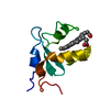
| Entry | Database: PDB / ID: 1h7m | ||||||
|---|---|---|---|---|---|---|---|
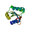
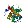
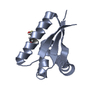
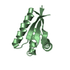
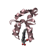
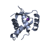
| Title | Ribosomal Protein L30e from Thermococcus celer | ||||||
 Components Components | 50S RIBOSOMAL PROTEIN L30E | ||||||
 Keywords Keywords | RIBOSOMAL PROTEIN / RNA-BINDING / RIBOSOME / THERMOPHILIC | ||||||
| Function / homology |  Function and homology information Function and homology informationcytosolic large ribosomal subunit / structural constituent of ribosome / translation / RNA binding Similarity search - Function | ||||||
| Biological species |   THERMOCOCCUS CELER (archaea) THERMOCOCCUS CELER (archaea) | ||||||
| Method |  X-RAY DIFFRACTION / MOLECULAR REPLACEMENT / Resolution: 1.96 Å X-RAY DIFFRACTION / MOLECULAR REPLACEMENT / Resolution: 1.96 Å | ||||||
 Authors Authors | Chen, Y.W. / Wong, K.B. | ||||||
 Citation Citation | Journal: Biochemistry / Year: 2003 Title: Crystal Structure of Ribosomal Protein L30E from the Extreme Thermophile Thermocccus Celer: Thermal Stability and RNA Binding Authors: Chen, Y.W. / Bycroft, M. / Wong, K.B. #1: Journal: Acta Crystallogr.,Sect.D / Year: 2001 Title: Crystallization and Preliminary Crystallographic Studies of a Ribosomal Protein L30E from the Hyperthermophilic Archaeon Thermococcus Celer Authors: Wong, K.B. / Wang, W.K. / Proctor, M.R. / Bycroft, M. / Chen, Y.W. #2: Journal: J.Mol.Biol. / Year: 1999Title: Local Folding Coupled to RNA Binding in the Yeast Ribosomal Protein L30 Authors: Mao, H. / Willamson, J.R. #3: Journal: Nat.Struct.Biol. / Year: 1999 Title: A Novel Loop-Loop Recognition Motif in the Yeast Ribosomal Protein L30 Autoregulatory RNA Complex Authors: Mao, H. / White, S.A. / Willamson, J.R. #4: Journal: Science / Year: 2000Title: The Complete Atomic Structure of the Large Ribosomal Subunit at 2.4 A Resolution Authors: Ban, N. / Nissen, P. / Hansen, J. / Moore, P.B. / Steitz, T.A. | ||||||
| History |
|
- Structure visualization
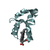
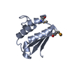
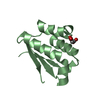
| Structure viewer | Molecule: MolmilJmol/JSmol |
|---|
- Downloads & links
-Download
| PDBx/mmCIF format | 1h7m.cif.gz | 33 KB | Display | PDBx/mmCIF format |
|---|---|---|---|---|
| PDB format | pdb1h7m.ent.gz | 22.3 KB | Display | PDB format |
| PDBx/mmJSON format | 1h7m.json.gz | Tree view | PDBx/mmJSON format | |
| Others |  Other downloads Other downloads |
-Validation report
| Arichive directory | https://data.pdbj.org/pub/pdb/validation_reports/h7/1h7mftp://data.pdbj.org/pub/pdb/validation_reports/h7/1h7m | HTTPS FTP |
|---|
-Related structure data
| Related structure data | |
|---|---|
| Similar structure data |


-Links
 PDBj
PDBj


- Assembly
| Deposited unit | 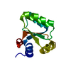
| ||||||||
|---|---|---|---|---|---|---|---|---|---|
| 1 |
| ||||||||
| Unit cell |
|
-Components
| #1: Protein | Mass: 10994.643 Da / Num. of mol.: 1 Source method: isolated from a genetically manipulated source Source: (gene. exp.) THERMOCOCCUS CELER (archaea) / Plasmid: PRSET-A / Production host:  |
|---|---|
| #2: Water | ChemComp-HOH /  Mass: 18.015 Da / Num. of mol.: 120 / Source method: isolated from a natural source / Formula: H2O Mass: 18.015 Da / Num. of mol.: 120 / Source method: isolated from a natural source / Formula: H2O |
| Compound details | BELONGS TO THE L30E FAMILY OF RIBOSOMAL PROTEINS. |
-Experimental details
-Experiment
| Experiment | Method: X-RAY DIFFRACTION / Number of used crystals: 1 |
|---|
- Sample preparation
| Crystal | Density Matthews: 2.68 Å3/Da / Density % sol: 54 % | ||||||||||||||||||||
|---|---|---|---|---|---|---|---|---|---|---|---|---|---|---|---|---|---|---|---|---|---|
| Crystal grow | pH: 5.6 / Details: 50MM KH2PO4, 20%(W/V) PEG8000, pH 5.60 | ||||||||||||||||||||
| Crystal grow | *PLUS Temperature: 290 K / Method: vapor diffusion, hanging dropDetails: Wong, K.B., (2001) Acta Crystallogr.,Sect.D, D57, 865. | ||||||||||||||||||||
| Components of the solutions | *PLUS
|
-Data collection
| Diffraction | Mean temperature: 100 K |
|---|---|
| Diffraction source | Source: ROTATING ANODE / Type: RIGAKU RU300 / Wavelength: 1.5418 |
| Detector | Type: MARRESEARCH / Detector: IMAGE PLATE / Date: Jul 17, 2000 / Details: MIRRORS |
| Radiation | Monochromator: NI FILTER / Protocol: SINGLE WAVELENGTH / Monochromatic (M) / Laue (L): M / Scattering type: x-ray |
| Radiation wavelength | Wavelength: 1.5418 Å / Relative weight: 1 |
| Reflection | Resolution: 1.96→24.2 Å / Num. obs: 8226 / % possible obs: 99.9 % / Observed criterion σ(I): 2 / Redundancy: 7.5 % / Rmerge(I) obs: 0.082 / Net I/σ(I): 7.6 |
| Reflection shell | Resolution: 1.96→2.07 Å / Redundancy: 7.3 % / Rmerge(I) obs: 0.343 / Mean I/σ(I) obs: 2.1 / % possible all: 99.9 |
- Processing
| Software |
| ||||||||||||||||||||
|---|---|---|---|---|---|---|---|---|---|---|---|---|---|---|---|---|---|---|---|---|---|
| Refinement | Method to determine structure: MOLECULAR REPLACEMENT Starting model: UNPUBLISHED NMR STRUCTURE OF THE SAME L30E PROTEIN Resolution: 1.96→24.18 Å / SU B: 3.612 / SU ML: 0.106 / Cross valid method: THROUGHOUT / ESU R: 0.15 / ESU R Free: 0.151 Details: GLY-97 - GLU-100 ARE NOT SEEN IN THE DENISTY MAPS SIDECHAINS OF MET-18, SER-67 AND SER-80 HAVE MULTIPLE CONFORMATIONS
| ||||||||||||||||||||
| Displacement parameters | Biso mean: 21.1 Å2
| ||||||||||||||||||||
| Refinement step | Cycle: LAST / Resolution: 1.96→24.18 Å
| ||||||||||||||||||||
| Refinement | *PLUS Lowest resolution: 24.2 Å / Rfactor obs: 0.17 / Rfactor Rwork: 0.17 | ||||||||||||||||||||
| Solvent computation | *PLUS | ||||||||||||||||||||
| Displacement parameters | *PLUS | ||||||||||||||||||||
| Refine LS restraints | *PLUS
|
