 Movie
Movie Controller


+ Open data

- Basic information
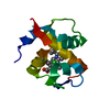
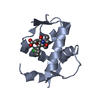
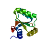
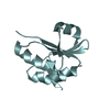
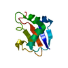
| Entry | Database: PDB / ID: 2cx6 | ||||||
|---|---|---|---|---|---|---|---|
| Title | Crystal structure of ribonuclease inhibitor Barstar | ||||||
 Components Components | Hypothetical protein yhcO | ||||||
 Keywords Keywords | HYDROLASE INHIBITOR / Barstar / ribonuclease inhibitor / RSGI / Structural Genomics / NPPSFA / National Project on Protein Structural and Functional Analyses / RIKEN Structural Genomics/Proteomics Initiative | ||||||
| Function / homology | Barstar-like / Barstar (barnase inhibitor) / Barstar (barnase inhibitor) / Barstar-like superfamily / Barnase; Chain D / 2-Layer Sandwich / Alpha Beta / Ribonuclease inhibitor barstar-like protein YhcO Function and homology information Function and homology information | ||||||
| Biological species |  | ||||||
| Method |  X-RAY DIFFRACTION / SYNCHROTRON / MAD / Resolution: 2.43 Å X-RAY DIFFRACTION / SYNCHROTRON / MAD / Resolution: 2.43 Å | ||||||
 Authors Authors | Murayama, K. / Kawazoe, M. / Shirouzu, M. / Yokoyama, S. / RIKEN Structural Genomics/Proteomics Initiative (RSGI) | ||||||
 Citation Citation | Journal: To be Published Title: Crystal structure of ribonuclease inhibitor Barstar Authors: Murayama, K. / Kawazoe, M. / Shirouzu, M. / Yokoyama, S. | ||||||
| History |
|
- Structure visualization
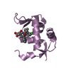
| Structure viewer | Molecule: MolmilJmol/JSmol |
|---|
- Downloads & links
-Download
| PDBx/mmCIF format | 2cx6.cif.gz | 50.1 KB | Display | PDBx/mmCIF format |
|---|---|---|---|---|
| PDB format | pdb2cx6.ent.gz | 36.6 KB | Display | PDB format |
| PDBx/mmJSON format | 2cx6.json.gz | Tree view | PDBx/mmJSON format | |
| Others |  Other downloads Other downloads |
-Validation report
| Arichive directory | https://data.pdbj.org/pub/pdb/validation_reports/cx/2cx6ftp://data.pdbj.org/pub/pdb/validation_reports/cx/2cx6 | HTTPS FTP |
|---|
-Related structure data
| Similar structure data | |
|---|---|
| Other databases |


-Links
 PDBj
PDBj- Assembly
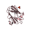
| Deposited unit | 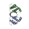
| ||||||||
|---|---|---|---|---|---|---|---|---|---|
| 1 | 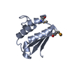
| ||||||||
| 2 | 
| ||||||||
| Unit cell |
|
-Components
| #1: Protein | Mass: 10902.902 Da / Num. of mol.: 2 Source method: isolated from a genetically manipulated source Source: (gene. exp.) #2: Water | ChemComp-HOH / |  Mass: 18.015 Da / Num. of mol.: 34 / Source method: isolated from a natural source / Formula: H2O Mass: 18.015 Da / Num. of mol.: 34 / Source method: isolated from a natural source / Formula: H2OHas protein modification | Y | |
|---|
-Experimental details
-Experiment
| Experiment | Method: X-RAY DIFFRACTION / Number of used crystals: 1 |
|---|
- Sample preparation
| Crystal | Density Matthews: 2.69 Å3/Da / Density % sol: 54.29 % |
|---|---|
| Crystal grow | Temperature: 293 K / Method: vapor diffusion, hanging drop / pH: 5.6 Details: 1.7M Sodium formate, 0.1M Sodium acetate, pH 5.6, VAPOR DIFFUSION, HANGING DROP, temperature 293K |
-Data collection
| Diffraction | Mean temperature: 100 K | ||||||||||||
|---|---|---|---|---|---|---|---|---|---|---|---|---|---|
| Diffraction source | Source: SYNCHROTRON / Site: SPring-8  / Beamline: BL26B2 / Wavelength: 0.9792, 0.9795, 0.964 / Beamline: BL26B2 / Wavelength: 0.9792, 0.9795, 0.964 | ||||||||||||
| Detector | Type: RIGAKU / Detector: IMAGE PLATE / Date: Jul 24, 2004 | ||||||||||||
| Radiation | Monochromator: Si 111 CHANNEL / Protocol: MAD / Monochromatic (M) / Laue (L): M / Scattering type: x-ray | ||||||||||||
| Radiation wavelength |
| ||||||||||||
| Reflection | Resolution: 2.43→50 Å / Num. obs: 9619 / % possible obs: 99.3 % / Observed criterion σ(I): -3 / Redundancy: 8.9 % / Biso Wilson estimate: 29.4 Å2 / Rsym value: 0.062 / Net I/σ(I): 27.7 | ||||||||||||
| Reflection shell | Resolution: 2.43→2.52 Å / Rsym value: 0.232 / % possible all: 100 |
- Processing
| Software |
| ||||||||||||||||||||||||||||||||||||
|---|---|---|---|---|---|---|---|---|---|---|---|---|---|---|---|---|---|---|---|---|---|---|---|---|---|---|---|---|---|---|---|---|---|---|---|---|---|
| Refinement | Method to determine structure: MAD / Resolution: 2.43→49.13 Å / Rfactor Rfree error: 0.009 / Data cutoff high absF: 1287230.85 / Data cutoff low absF: 0 / Isotropic thermal model: RESTRAINED / Cross valid method: THROUGHOUT / σ(F): 0
| ||||||||||||||||||||||||||||||||||||
| Solvent computation | Solvent model: FLAT MODEL / Bsol: 36.8107 Å2 / ksol: 0.362069 e/Å3 | ||||||||||||||||||||||||||||||||||||
| Displacement parameters | Biso mean: 45.5 Å2
| ||||||||||||||||||||||||||||||||||||
| Refine analyze |
| ||||||||||||||||||||||||||||||||||||
| Refinement step | Cycle: LAST / Resolution: 2.43→49.13 Å
| ||||||||||||||||||||||||||||||||||||
| Refine LS restraints |
| ||||||||||||||||||||||||||||||||||||
| LS refinement shell | Resolution: 2.43→2.58 Å / Rfactor Rfree error: 0.035 / Total num. of bins used: 6
| ||||||||||||||||||||||||||||||||||||
| Xplor file |
|
